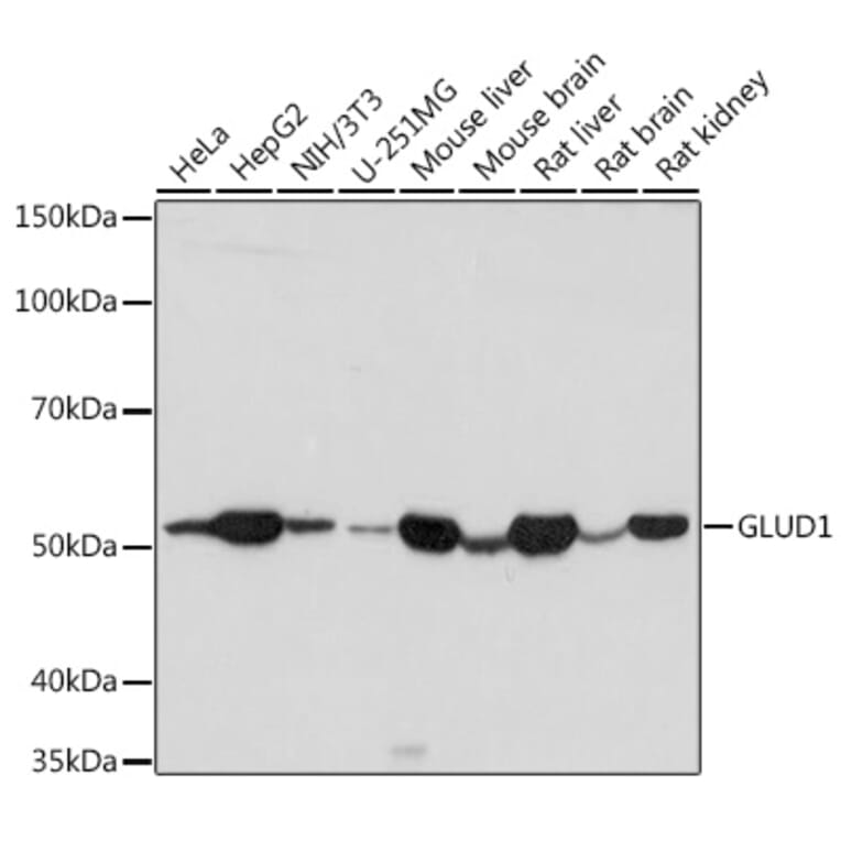
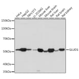
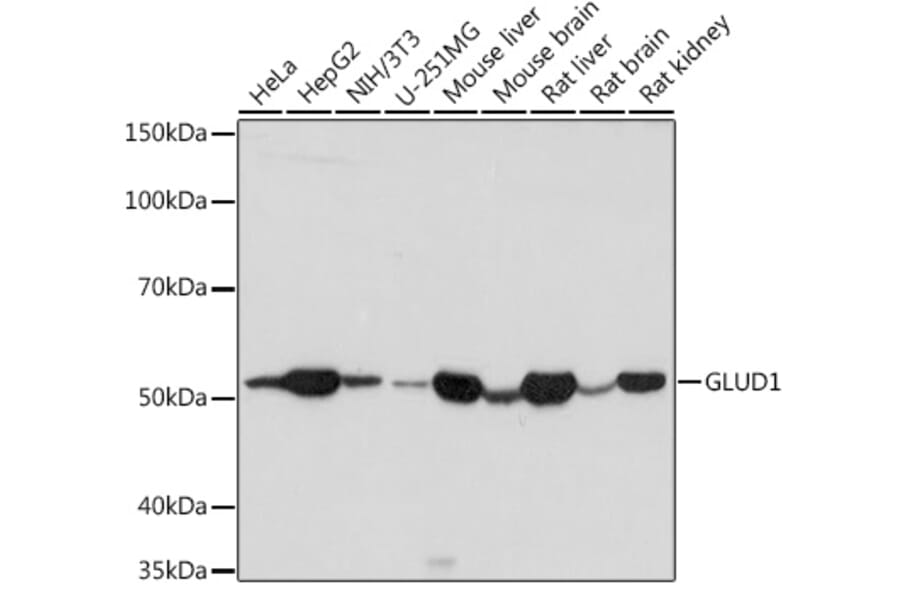
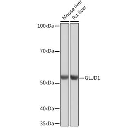
Western blot analysis of extracts of various cell lines, using Anti-GLUD1 Antibody (A15814) at 1:1,000 dilution. The secondary antibody was Goat Anti-Rabbit IgG H&L Antibody (HRP) at 1:10,000 dilution. Lysates/proteins were present at 25µg per lane. The blocking buffer used was 3% non-fat dry milk in TBST. Detection was with a ECL Basic Kit. Exposure time: 10s.
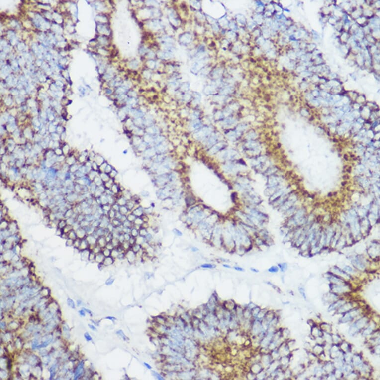
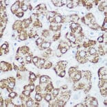
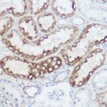
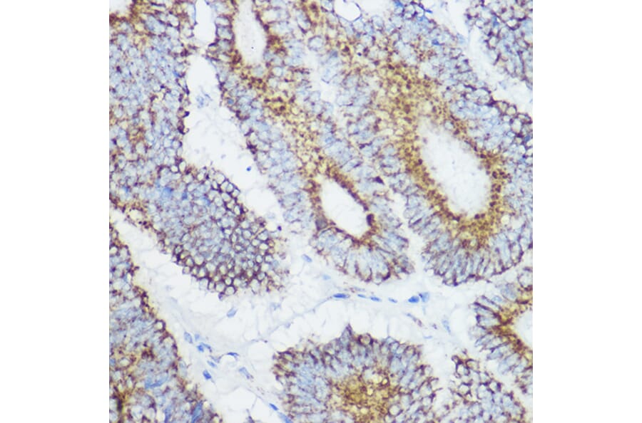
Immunohistochemistry analysis of paraffin-embedded human colon carcinoma tissue using Anti-GLUD1 Antibody (A15814) at a dilution of 1:100 (40x lens). Perform high pressure antigen retrieval with 10 mM citrate buffer pH 6.0 before commencing with IHC staining protocol.
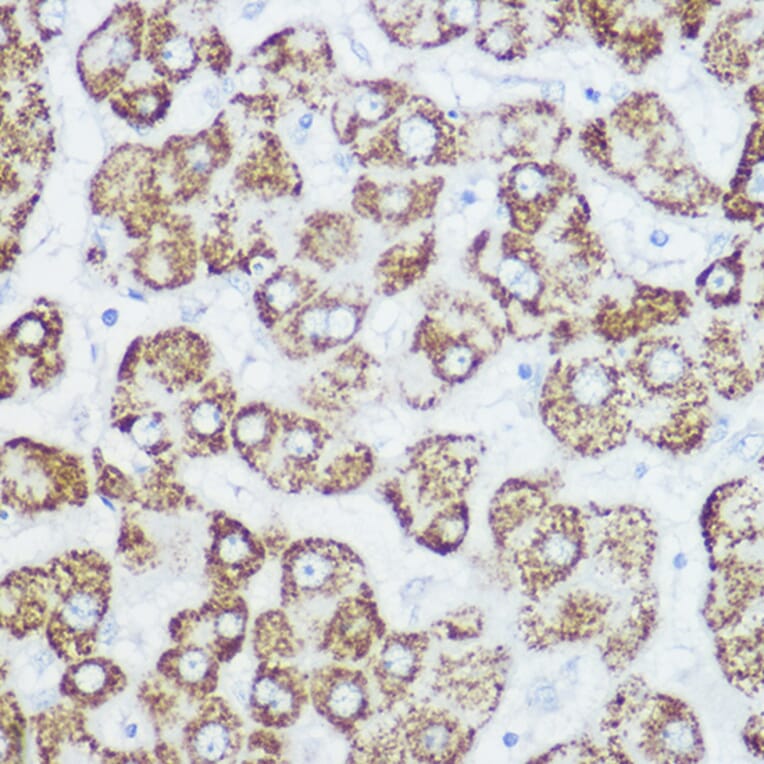
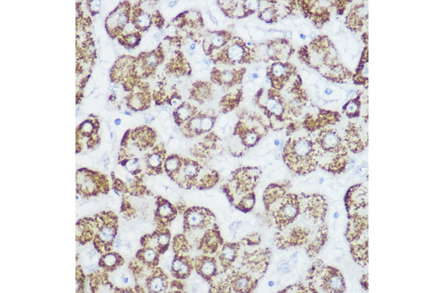
Immunohistochemistry analysis of paraffin-embedded human liver using Anti-GLUD1 Antibody (A15814) at a dilution of 1:100 (40x lens). Perform high pressure antigen retrieval with 10 mM citrate buffer pH 6.0 before commencing with IHC staining protocol.
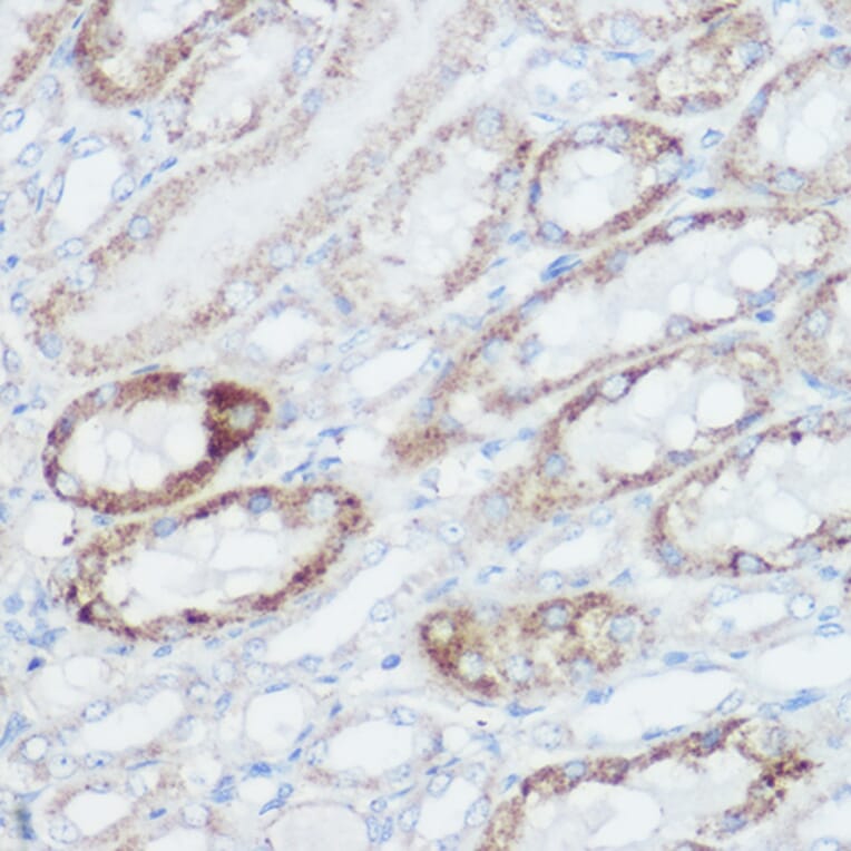
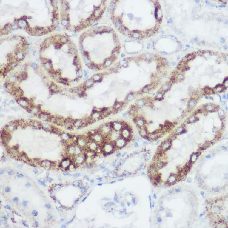
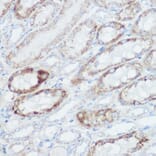
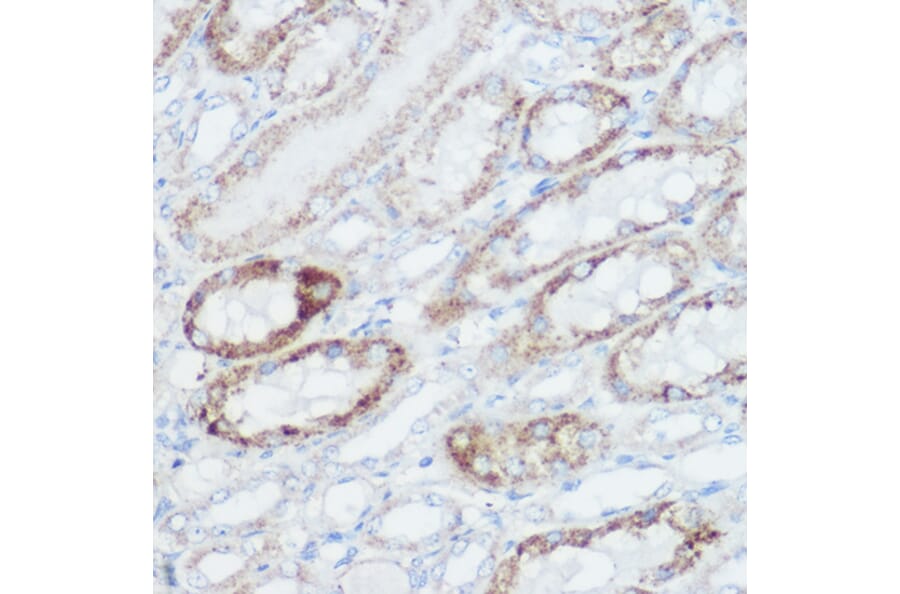
Immunohistochemistry analysis of paraffin-embedded mouse kidney using Anti-GLUD1 Antibody (A15814) at a dilution of 1:100 (40x lens). Perform high pressure antigen retrieval with 10 mM citrate buffer pH 6.0 before commencing with IHC staining protocol.
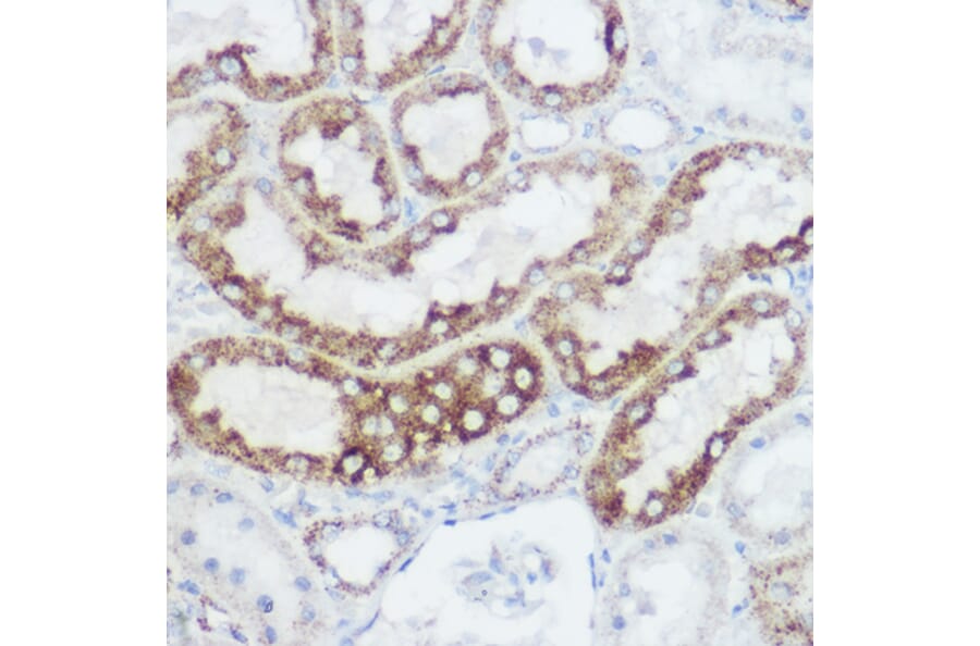
Immunohistochemistry analysis of paraffin-embedded rat kidney using Anti-GLUD1 Antibody (A15814) at a dilution of 1:100 (40x lens). Perform high pressure antigen retrieval with 10 mM citrate buffer pH 6.0 before commencing with IHC staining protocol.
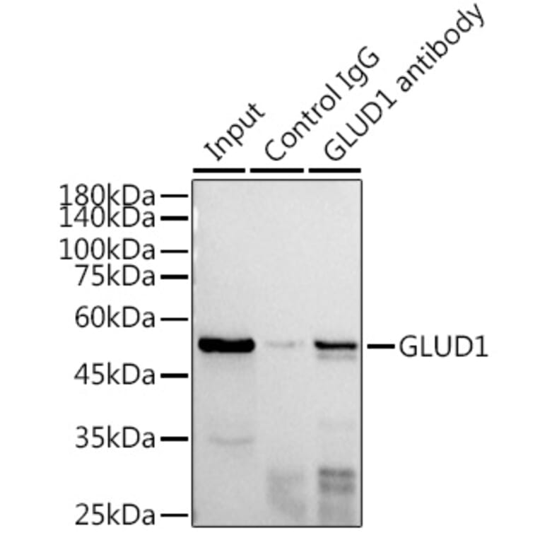
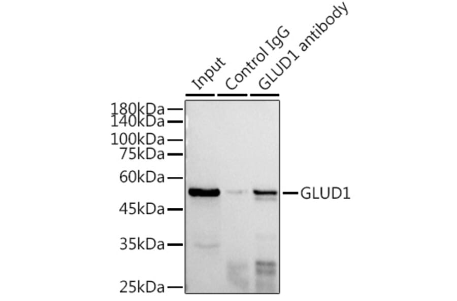
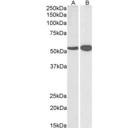
Immunoprecipitation analysis of 300µg extracts of HepG2 cells using 3µg of Anti-GLUD1 Antibody (A15814). This Western blot was performed on the immunoprecipitate using Anti-GLUD1 Antibody (A15814) at a dilution of 1:1000.