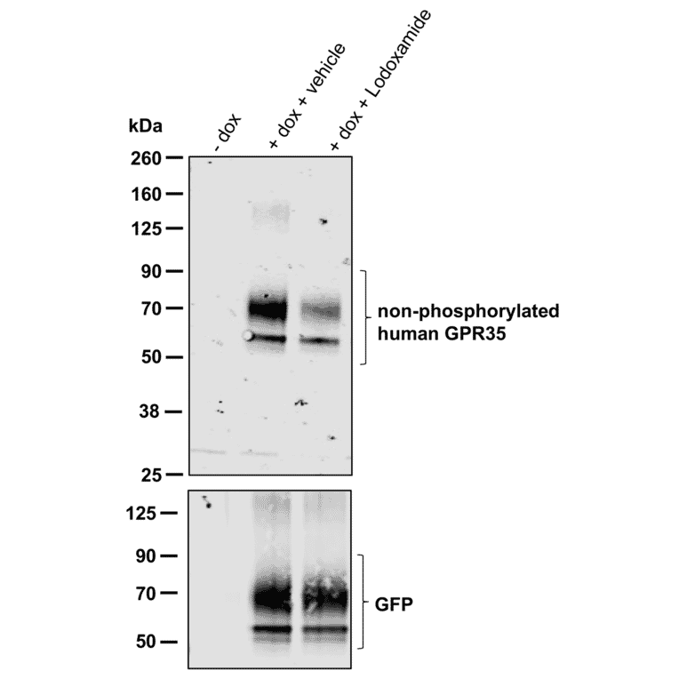
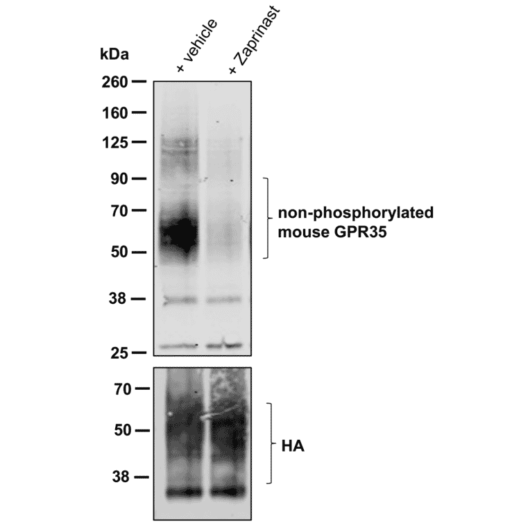
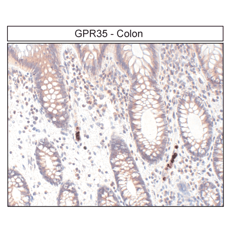
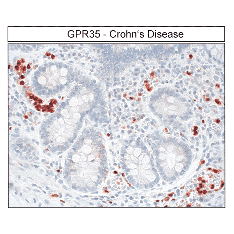
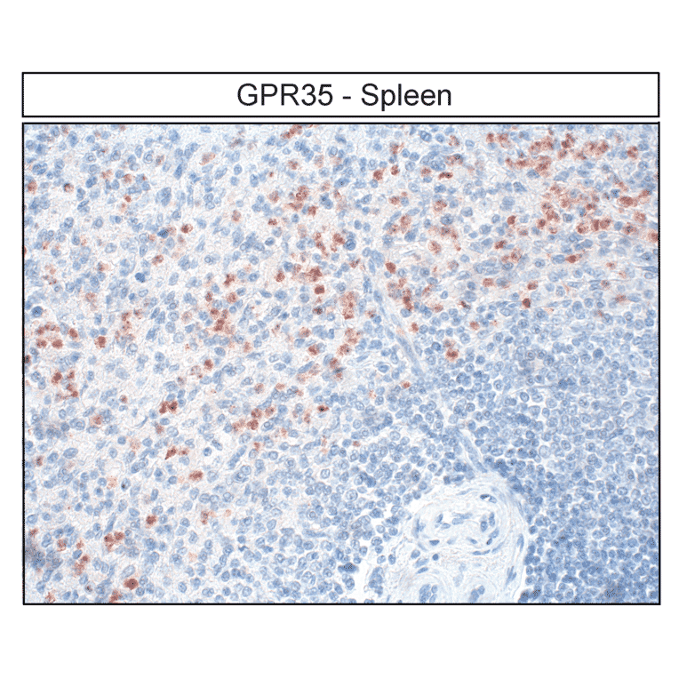
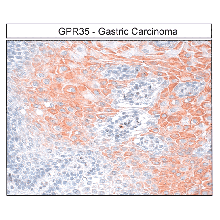
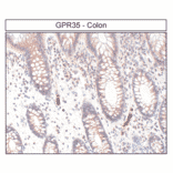
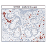
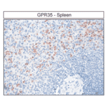
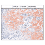
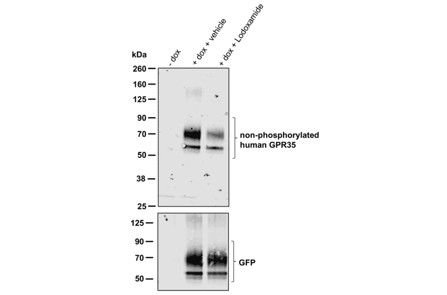
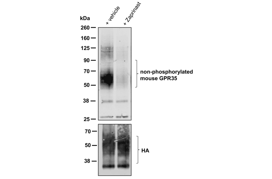
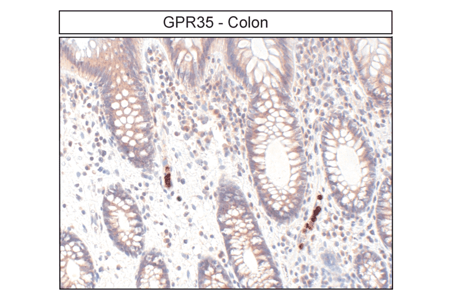
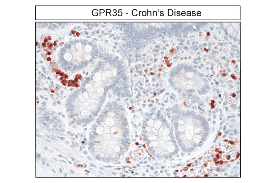
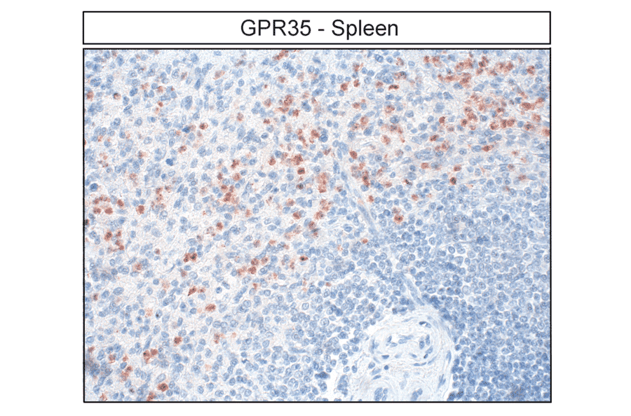
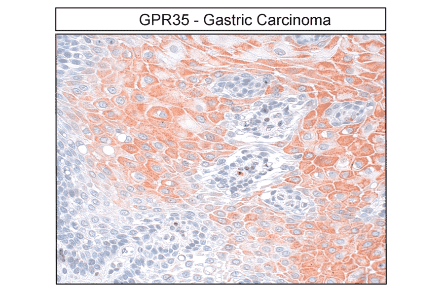
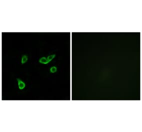
This antibody binds specifically to the C-terminal domain of human GPR35, detecting the non-phosphorylated form. It is suitable for Western blot analysis and supports immunohistochemistry in formalin-fixed, paraffin-embedded tissue sections.
Applications
WB, IHC
Dilutions
WB: 1,000, IHC: 1:100
Reactivity
Human
Immunogen
Synthetic peptide corresponding to the C-terminal of human GPR35. Immunogen range is 22-22 amino acids.
Host
Rabbit
Clonality
Polyclonal
Isotype
IgG
Conjugate
Unconjugated
Purification
Antigen affinity purification.
Concentration
Lot Specific
Product Form
Liquid
Formulation
Supplied in Dulbecco's PBS, pH 7.4, with 150 mM NaCl and 0.005% Sodium Azide.
Storage
Shipped at 4°C. Upon delivery aliquot and store at -20°C. Avoid freeze/thaw cycles.
Detection of non-phosphorylated human GPR35 Receptor (GPR35) by Western blot. Flp-In TREx 293 cells harboring human GPR35-eYFP, induced to express the receptor, were treated with vehicle or 100 nM Lodoxamide for 5 min. Lysates were enriched with a GFP-trap and immunoblotted with anti-GPR35 antibody (A334529) at 1:1000 (upper panel) or anti-GFP at 1:10,000 to confirm expression and equal loading (lower panel).
Western blot analysis of non-phosphorylated mouse GPR35 Receptor (GPR35). Flp-In TREx 293 cells harboring mouse GPR35-HA, induced to express the receptor, were treated with vehicle or 10 µM Zaprinast for 5 min. Lysates were enriched with an HA-trap and probed with anti-GPR35 antibody (A334529) at 1:1000 (upper panel) or anti-HA at 1:10,000 to confirm expression and equal loading (lower panel).
Immunohistochemical localization of GPR35 Receptor (GPR35) in human colon. Colon sections were dewaxed, microwaved in citric acid, and stained with anti-GPR35 antibody (A334529) at 1:100, followed by biotinylated anti-rabbit IgG and avidin-biotin solution. Color was developed with 3-amino-9-ethylcarbazole (AEC), and sections were counterstained with hematoxylin. GPR35 receptors were detected at the plasma membrane of epithelial cells and macrophages.
Detection of GPR35 Receptor (GPR35) in Crohn’s disease by immunohistochemistry. Colon sections were dewaxed, microwaved in citric acid, and incubated with anti-GPR35 antibody (A334529) at 1:100, followed by biotinylated anti-rabbit IgG and avidin-biotin solution. Color was developed with 3-amino-9-ethylcarbazole (AEC), and sections were counterstained with hematoxylin. GPR35 receptors were observed at the plasma membrane of epithelial cells and macrophages.
Immunohistochemical detection of GPR35 Receptor (GPR35) in human spleen. Spleen sections were dewaxed, microwaved in citric acid, and stained with anti-GPR35 antibody (A334529) at 1:100, followed by biotinylated anti-rabbit IgG and avidin-biotin solution. Color was developed with 3-amino-9-ethylcarbazole (AEC), and sections were counterstained with hematoxylin. GPR35 receptors were detected in macrophages.
Localization of GPR35 Receptor (GPR35) in gastric carcinoma by immunohistochemistry. Tumor sections were dewaxed, microwaved in citric acid, and incubated with anti-GPR35 antibody (A334529) at 1:100, followed by biotinylated anti-rabbit IgG and avidin-biotin solution. Color was developed with 3-amino-9-ethylcarbazole (AEC), and sections were counterstained with hematoxylin. GPR35 receptors were observed at the plasma membrane of tumor cells.