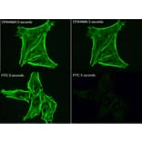
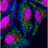
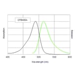
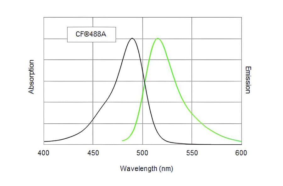
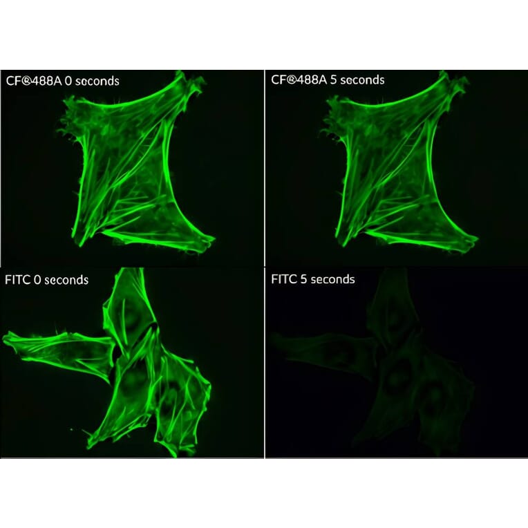
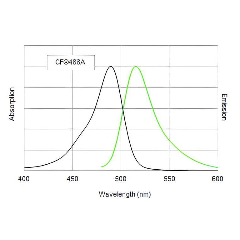
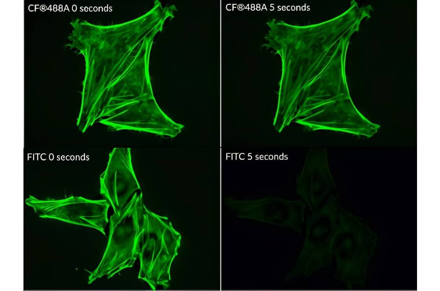
This polyclonal rabbit anti-HA antibody was raised against the hemagglutinin (HA) epitope tag and specifically recognizes the YPYDVPDYA sequence.
Applications
ICC/IF, IHC, WB, Flow Cytometry
Dilutions
IF: 1-10 µg/mL, WB: 50-100 ng/mL, IHC: 1-10 µg/mL
Reactivity
Species Independent
Host
Rabbit
Clonality
Polyclonal
Isotype
IgG
Conjugate
CF®488A
Concentration
2 mg/mL
Product Form
Liquid
Reconstitution
CF® Dye and biotin conjugates: Add 0.5 ml dH2O for reconstitution. HRP or DNP conjugates: Add 1 ml dH2O for reconstitution.
Formulation
Supplied in phosphate buffered saline (PBS) containing 50% glycerol, 2 mg/ml BSA, and 0.05% sodium azide
Storage
Shipped at +4°C. Upon delivery aliquot and store at -20°C. Avoid freeze/thaw cycles. This product is also photosensitive and should be protected from light. Should this product contain a precipitate we recommend microcentrifugation before use. CF® Dyes are guaranteed for at least 6 months from data of receipt when stored correctly.
General Notes
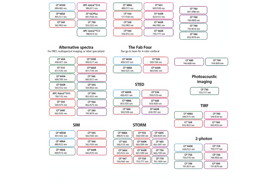
Looking for a specific protein conjugate to simplify your workflow? We offer a library of over 2,000 targets conjugated to your choice of CF® dye. To enquire about a custom product, contact us directly.
Disclaimer
This product is for research use only. It is not intended for diagnostic or therapeutic use.
CF®488A exhibits substantially greater photostability than FITC. HeLa cells were stained with either CF®488A phalloidin or FITC phalloidin and imaged at time zero or after 5 seconds of continuous excitation using a FITC filter set. FITC fluorescence photobleaches almost immediately, whereas CF®488A retains strong signal intensity, demonstrating its high photostability.
This chart summarizes commonly used CF® dyes grouped by their suitability for specific imaging modalities, including alternative spectra applications, four-color confocal imaging, near-infrared western blotting, photoacoustic imaging, STED, SIM, STORM, TIRF, and two-photon microscopy. Each dye is shown with its characteristic excitation and emission wavelengths (nm), providing a practical reference for selecting spectrally compatible dyes and optimizing multicolor experimental design across a range of fluorescence techniques.
HeLa cells were immunostained with rabbit anti-COX IV followed by CF®488A–conjugated goat anti-rabbit secondary antibodies to label mitochondria in green, and with mouse anti-Golgin 97 followed by CF®555–conjugated goat anti-mouse secondary antibodies to label the Golgi apparatus in red. Actin filaments were visualized using CF®405-conjugated phalloidin, producing a blue signal, and cell nuclei were counterstained with RedDot™2, appearing magenta.
Normalized emission spectra of the CF® dye family spanning the visible to near-infrared range are shown, illustrating the spectral diversity and overlap between dyes. Curves represent relative fluorescence intensity as a function of emission wavelength (nm), with peak positions corresponding to each dye’s characteristic emission maximum. This reference highlights the broad coverage of CF® dyes for multicolor fluorescence applications and aids in selecting compatible dye combinations for imaging, flow cytometry, and other fluorescence-based assays.