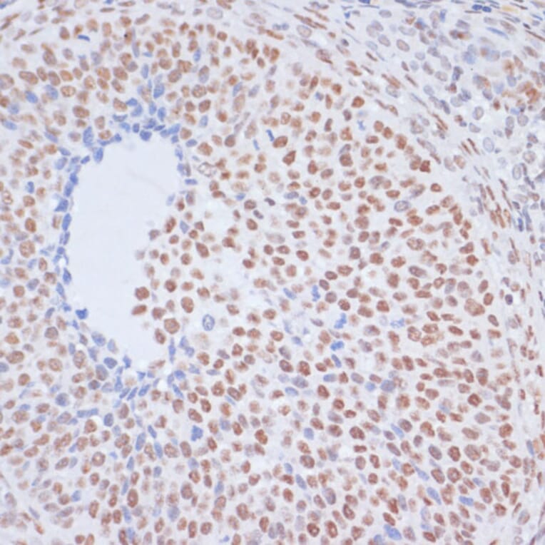
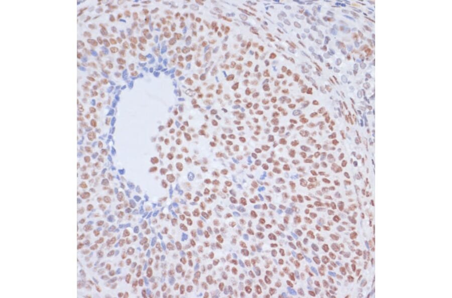
Immunohistochemistry analysis of paraffin-embedded rat ovary using Anti-Histone H1.4 (acetyl Lys26) Antibody (A305416) at a dilution of 1:200 (40x lens). Perform microwave antigen retrieval with 10 mM PBS buffer pH 7.2 before commencing with IHC staining protocol.
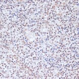
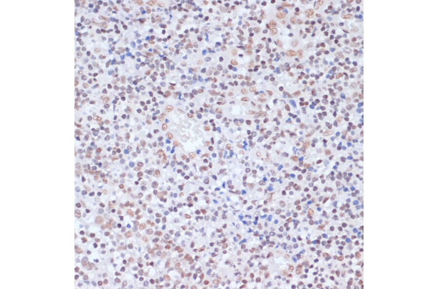
Immunohistochemistry analysis of paraffin-embedded human tonsil using Anti-Histone H1.4 (acetyl Lys26) Antibody (A305416) at a dilution of 1:200 (40x lens). Perform microwave antigen retrieval with 10 mM PBS buffer pH 7.2 before commencing with IHC staining protocol.
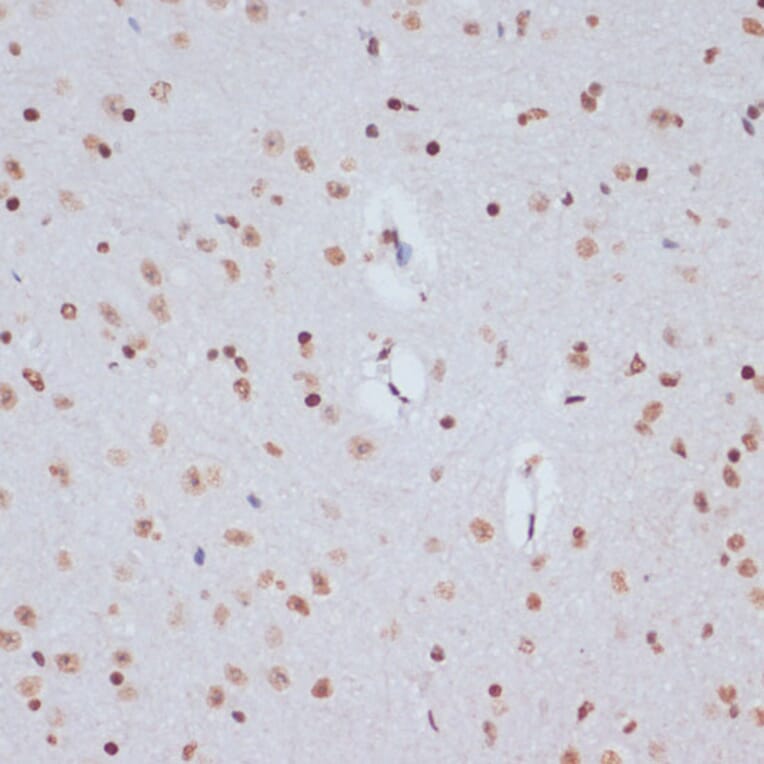
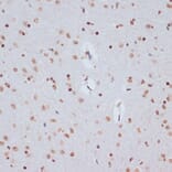
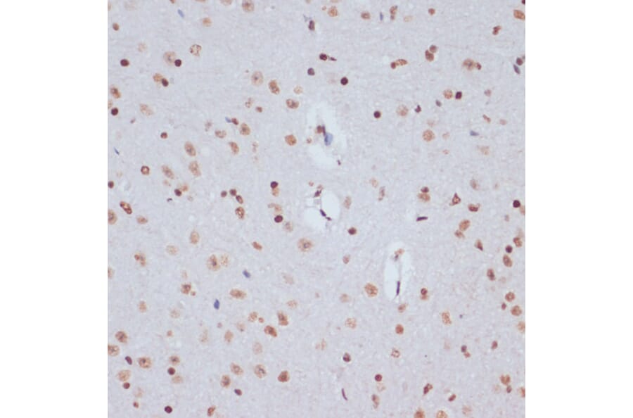
Immunohistochemistry analysis of paraffin-embedded mouse brain using Anti-Histone H1.4 (acetyl Lys26) Antibody (A305416) at a dilution of 1:200 (40x lens). Perform microwave antigen retrieval with 10 mM PBS buffer pH 7.2 before commencing with IHC staining protocol.
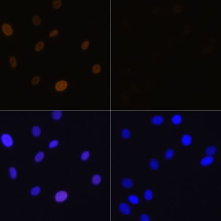
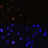
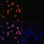
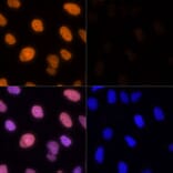
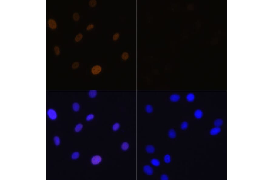
Immunofluorescence analysis of C6 cells using Anti-Histone H1.4 (acetyl Lys26) Antibody (A305416) at a dilution of 1:100. DAPI was used to stain the cell nuclei (blue). C6 cells were treated by TSA (1 uM) at 37°C for 18 hours. DAPI was used to stain the cell nuclei (blue).
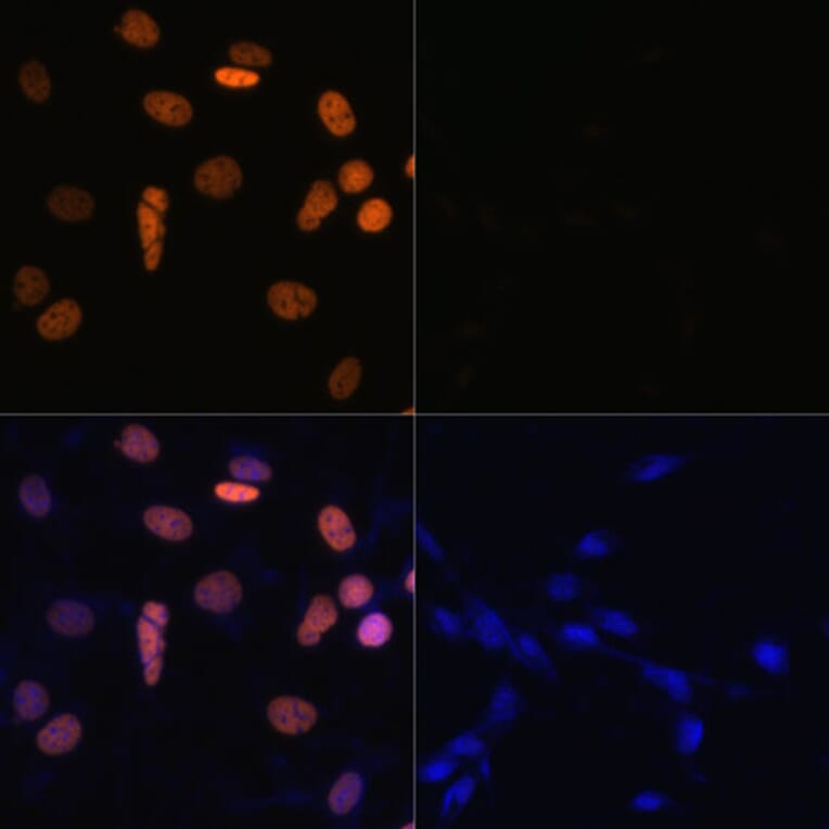
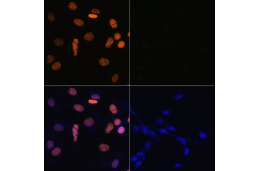
Immunofluorescence analysis of NIH/3T3 cells using Anti-Histone H1.4 (acetyl Lys26) Antibody (A305416) at a dilution of 1:100. DAPI was used to stain the cell nuclei (blue). NIH/3T3 cells were treated by TSA (1 uM) at 37°C for 18 hours. DAPI was used to stain the cell nuclei (blue).
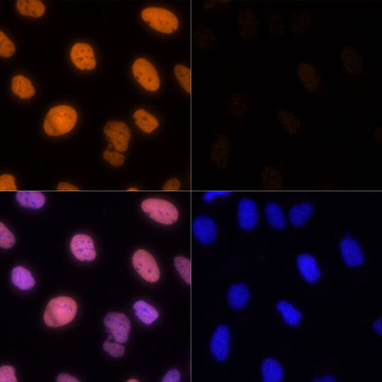
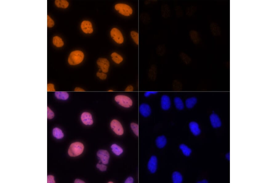
Immunofluorescence analysis of U-2 OS cells using Anti-Histone H1.4 (acetyl Lys26) Antibody (A305416) at a dilution of 1:100. DAPI was used to stain the cell nuclei (blue). U2OS cells were treated by TSA (1 uM) at 37°C for 18 hours. DAPI was used to stain the cell nuclei (blue).