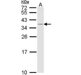
This antibody specifically binds to the empty but not peptide-loaded form of HLA-DR1. DR is the isotypes of human MHC Class II molecules expressed on antigen-presenting cells (i.e. APC: dendritic cells, B lymphocytes, monocytes, macrophages).
Applications
WB, Flow Cytometry, ELISA
Dilutions
Flow Cytometry: Stains immature dendritic cells that express empty cell surface MHC molecules, but not cells that express predominantly peptide loaded forms.
Reactivity
Human
Immunogen
Purified, insoluble DR1 beta chain (DRB1*0101) expressed in E. coli inclusion bodies.
Host
Mouse
Clonality
Monoclonal
Clone ID
MEM-267
Isotype
IgG2b
Conjugate
Unconjugated
Purification
Protein A chromatography.
Concentration
1 mg/ml
Product Form
Liquid
Formulation
Supplied in Phosphate Buffered Saline, pH 7.4, with 15 mM Sodium Azide.
Storage
Shipped at 4°C. Upon delivery aliquot and store at -20°C. Avoid freeze/thaw cycles.
Synonyms
HLA class II histocompatibility antigen, DR alpha chain, HLA-DRA, HLA-DRA1, MHC class II antigen DRA
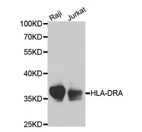
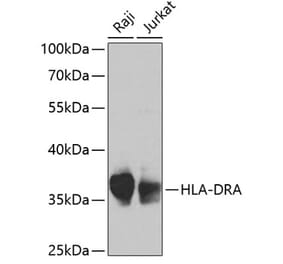
Anti-HLA-DR Antibody [MEM-267] (A85933) works in WB application under reducing and non-reducing conditions. Western blotting analysis was performed on whole cell extracts (RIPA lysis buffer) of Raji and Jurkat cell lines, mixed and heated (100°C, 5 min) with reducing and non-reducing SDS-loading buffer. Samples were resolved using 10% Tris-glycine SDS gel electrophoresis. Nitrocellulose membrane blot was probed with mouse IgG2b monoclonal antibody MEM-267 (1 µg/ml), followed by IRDye 800CW Goat-anti-Mouse IgG (green). Multiplex fluorescent Western blot detection was performed. HLA-DR1 molecules were detected at ~25 kDa in Raji cell line.
Separation of human HLA-DR1 positive lymphocytes (red-filled) from HLA-DR1 negative lymphocytes (black-dashed) in flow cytometry surface staining of peripheral whole blood, labeling HLA-DR with Anti-HLA-DR Antibody [MEM-267] (A85933), (concentration in sample 9 µg/ml, GAM APC).


![WB - Anti-HLA-DR Antibody [MEM-267] (A85933)](https://cdn.antibodies.com/image/catalog/85/A85933_1.jpg?profile=product_top)
![Flow Cytometry - Anti-HLA-DR Antibody [MEM-267] (A85933)](https://cdn.antibodies.com/image/catalog/85/A85933_2.jpg?profile=product_top)
![Flow Cytometry - Anti-HLA-DR Antibody [MEM-267] (A85933)](https://cdn.antibodies.com/image/catalog/85/A85933_3.jpg?profile=product_top)
![WB - Anti-HLA-DR Antibody [MEM-267] (A85933)](https://cdn.antibodies.com/image/catalog/85/A85933_1.jpg?profile=product_top_thumb)
![Flow Cytometry - Anti-HLA-DR Antibody [MEM-267] (A85933)](https://cdn.antibodies.com/image/catalog/85/A85933_2.jpg?profile=product_top_thumb)
![Flow Cytometry - Anti-HLA-DR Antibody [MEM-267] (A85933)](https://cdn.antibodies.com/image/catalog/85/A85933_3.jpg?profile=product_top_thumb)
![WB - Anti-HLA-DR Antibody [MEM-267] (A85933)](https://cdn.antibodies.com/image/catalog/85/A85933_1.jpg?profile=product_image)
![Flow Cytometry - Anti-HLA-DR Antibody [MEM-267] (A85933)](https://cdn.antibodies.com/image/catalog/85/A85933_2.jpg?profile=product_image)
![Flow Cytometry - Anti-HLA-DR Antibody [MEM-267] (A85933)](https://cdn.antibodies.com/image/catalog/85/A85933_3.jpg?profile=product_image)
![Flow Cytometry - Anti-HLA-DR Antibody [L243] (A86736)](https://cdn.antibodies.com/image/catalog/86/A86736_1.jpg?profile=product_alternative)
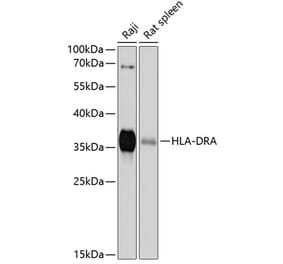
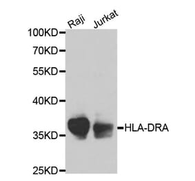
![SDS-PAGE - Anti-HLA-DR Antibody [IMMU-114] - Low endotoxin, Azide free (A324062) - Antibodies.com](https://cdn.antibodies.com/image/catalog/324/A324062_1.jpg?profile=product_alternative)
![Flow Cytometry - Anti-HLA-DR Antibody [MEM-12] (A85815)](https://cdn.antibodies.com/image/catalog/85/A85815_1.jpg?profile=product_alternative)
![Bioactivity-Binding - Anti-HLA-DR Antibody [1D09C3] - BSA and Azide free (A344134) - Antibodies.com](https://cdn.antibodies.com/image/catalog/344/A344134_1.jpg?profile=product_alternative)
![Flow Cytometry - Anti-HLA-DR Antibody [L243] - BSA and Azide free (A344135) - Antibodies.com](https://cdn.antibodies.com/image/catalog/344/A344135_1.jpg?profile=product_alternative)