Supplied in Phosphate Buffered Saline, pH 7.3, with 50% Glycerol and 0.05% Proclin 300.
Storage
Shipped at 4°C. Upon delivery aliquot and store at -20°C. Avoid freeze/thaw cycles.
Synonyms
ARE binding protein AUFI type A, AU-rich element RNA-binding protein 1, AUF, AUF1, AUF1A, Heterogeneous nuclear ribonucleoprotein D0, hnRNP D, hnRNP D0, Hnrnpd, hnRNPD0, HNRPD, HNRPD_HUMAN, P37
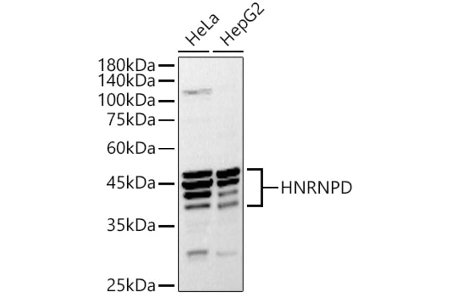
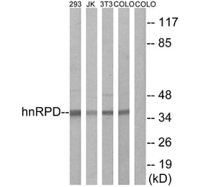
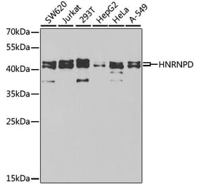
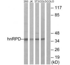
Western Blot - Anti-hnRNP D/AUF1 Antibody (A87646)
Western blot analysis of extracts of various cell lines, using Anti-hnRNP D/AUF1 Antibody (A87646) at 1:1,000 dilution. The secondary antibody was Goat Anti-Rabbit IgG H&L Antibody (HRP) at 1:10,000 dilution. Lysates/proteins were present at 25µg per lane. The blocking buffer used was 3% non-fat dry milk in TBST. Detection was with a ECL Basic Kit. Exposure time: 30s.
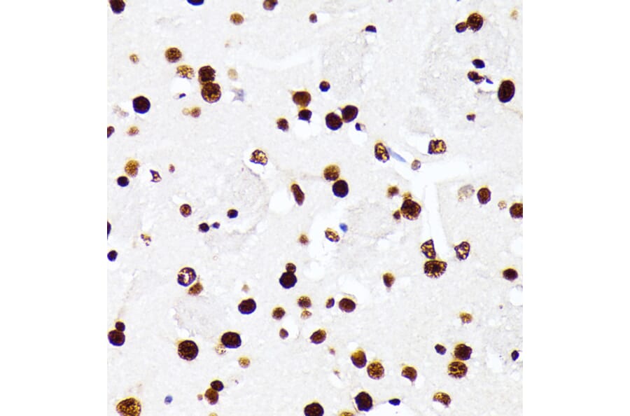
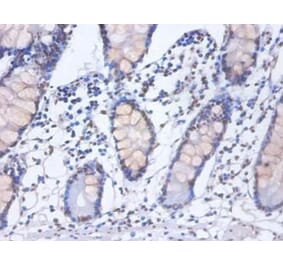
Immunohistochemistry analysis of paraffin-embedded rat liver using Anti-hnRNP D/AUF1 Antibody (A87646) at a dilution of 1:100 (40x lens). Perform microwave antigen retrieval with 10 mM PBS buffer pH 7.2 before commencing with IHC staining protocol.
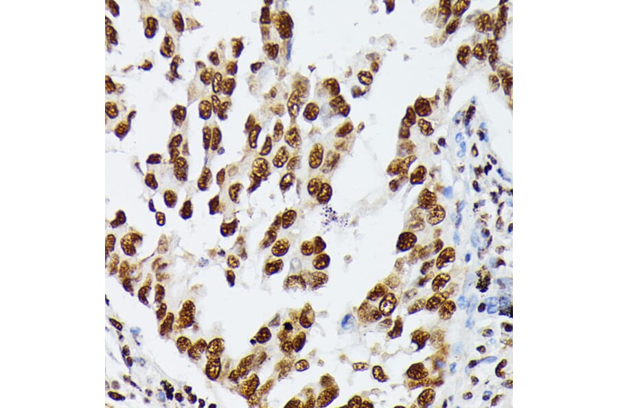
Immunohistochemistry analysis of paraffin-embedded human lung cancer using Anti-hnRNP D/AUF1 Antibody (A87646) at a dilution of 1:100 (40x lens). Perform microwave antigen retrieval with 10 mM PBS buffer pH 7.2 before commencing with IHC staining protocol.
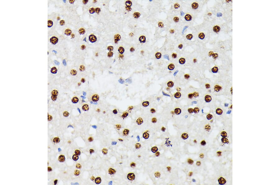
Immunohistochemistry analysis of paraffin-embedded mouse brain using Anti-hnRNP D/AUF1 Antibody (A87646) at a dilution of 1:100 (40x lens). Perform microwave antigen retrieval with 10 mM PBS buffer pH 7.2 before commencing with IHC staining protocol.
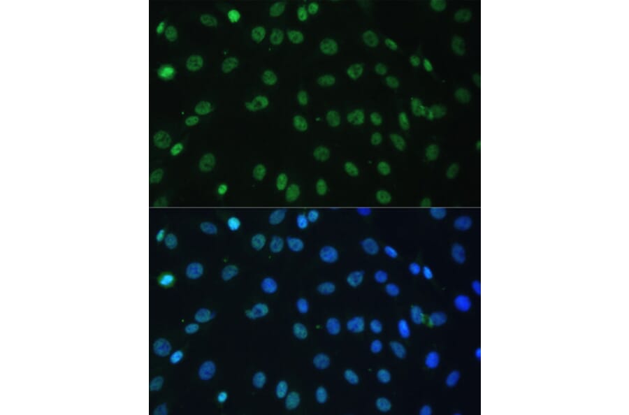
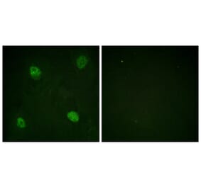
Immunofluorescence analysis of C6 cells using Anti-hnRNP D/AUF1 Antibody (A87646) at a dilution of 1:100. DAPI was used to stain the cell nuclei (blue).
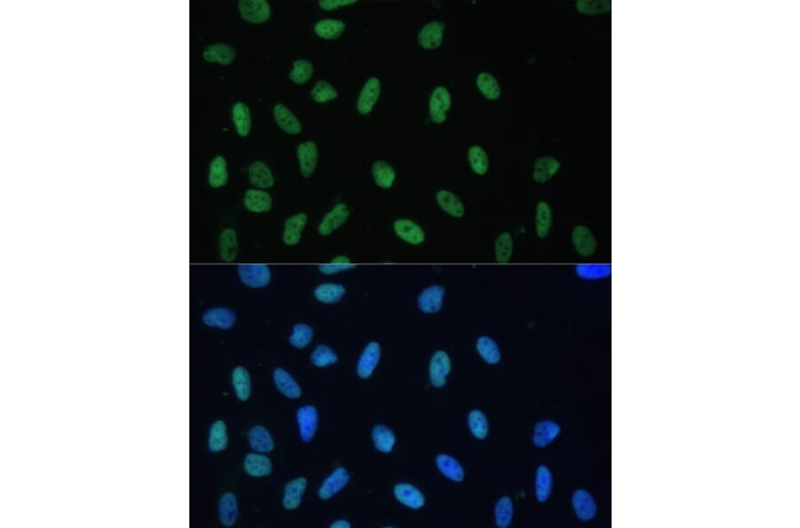
Immunofluorescence analysis of U-2 OS cells using Anti-hnRNP D/AUF1 Antibody (A87646) at a dilution of 1:100. DAPI was used to stain the cell nuclei (blue).
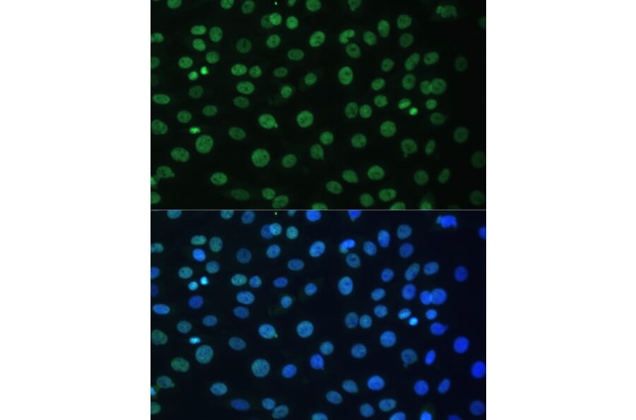
Immunofluorescence analysis of C6 cells using Anti-hnRNP D/AUF1 Antibody (A87646) at a dilution of 1:100. DAPI was used to stain the cell nuclei (blue).
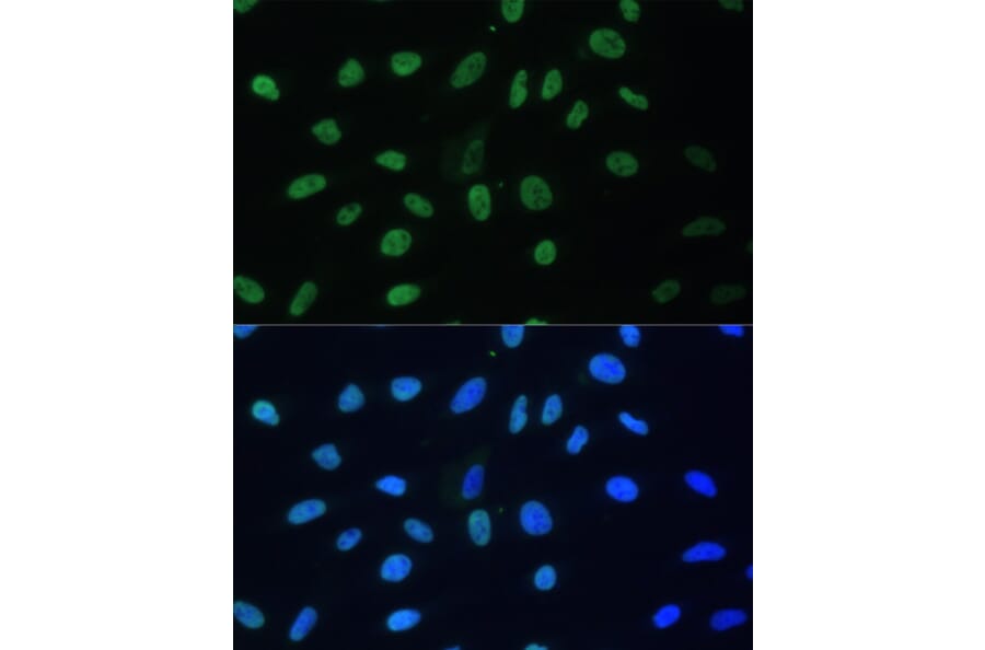
Immunofluorescence analysis of U-2 OS cells using Anti-hnRNP D/AUF1 Antibody (A87646) at a dilution of 1:100. DAPI was used to stain the cell nuclei (blue).
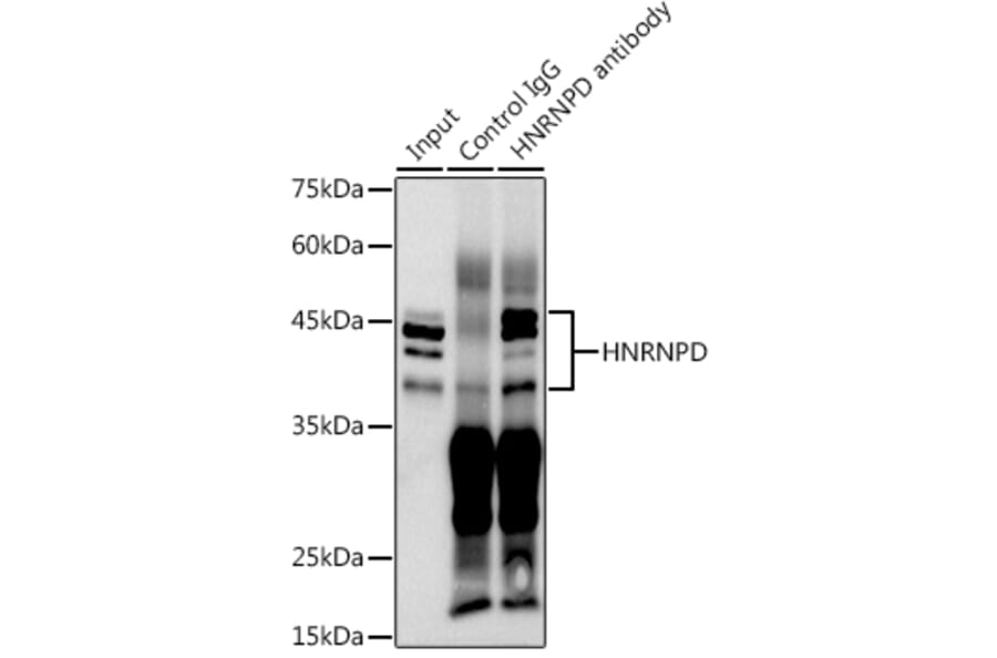
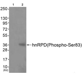
Western Blot - Anti-hnRNP D/AUF1 Antibody (A87646)
Immunoprecipitation analysis of 300µg extracts of HeLa cells using 3µg of Anti-hnRNP D/AUF1 Antibody (A87646). This Western blot was performed on the immunoprecipitate using Anti-hnRNP D/AUF1 Antibody (A87646) at a dilution of 1:1000.