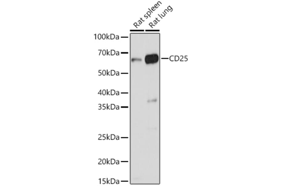
Western Blot - Anti-IL-2 Receptor alpha Antibody (A14290)
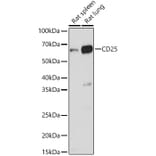
Western blot analysis of extracts of various cell lines, using Anti-IL-2 Receptor alpha Antibody (A14290) at 1:1,000 dilution. The secondary antibody was Goat Anti-Rabbit IgG H&L Antibody (HRP) at 1:10,000 dilution. Lysates/proteins were present at 25µg per lane. The blocking buffer used was 3% non-fat dry milk in TBST. Detection was with a ECL Basic Kit. Exposure time: 3s.
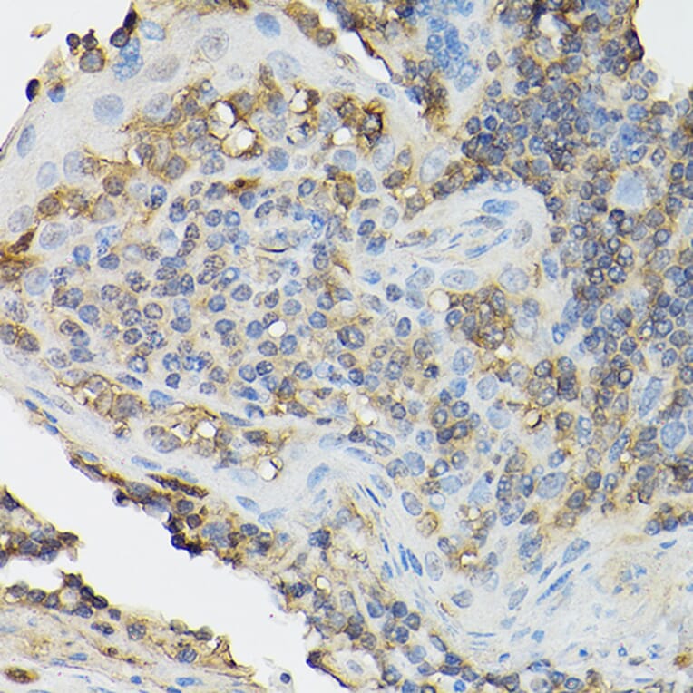
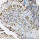
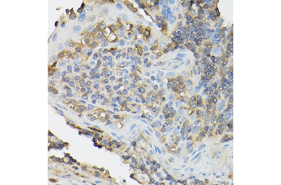
Immunohistochemistry analysis of paraffin-embedded human tonsil using Anti-IL-2 Receptor alpha Antibody (A14290) at a dilution of 1:100 (40x lens). Perform high pressure antigen retrieval with 10 mM citrate buffer pH 6.0 before commencing with IHC staining protocol.
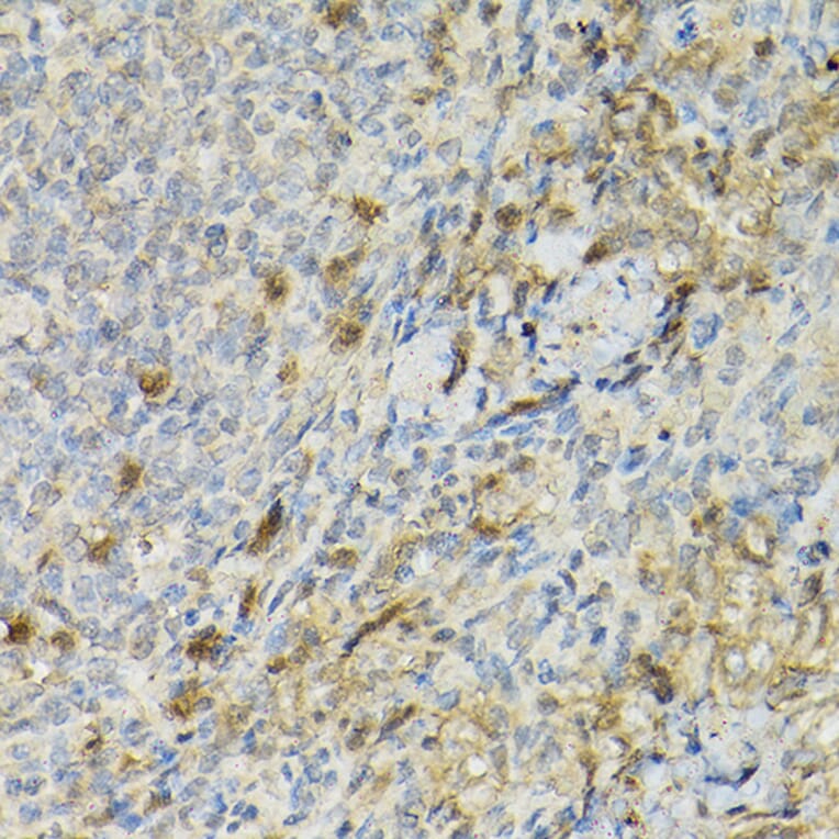
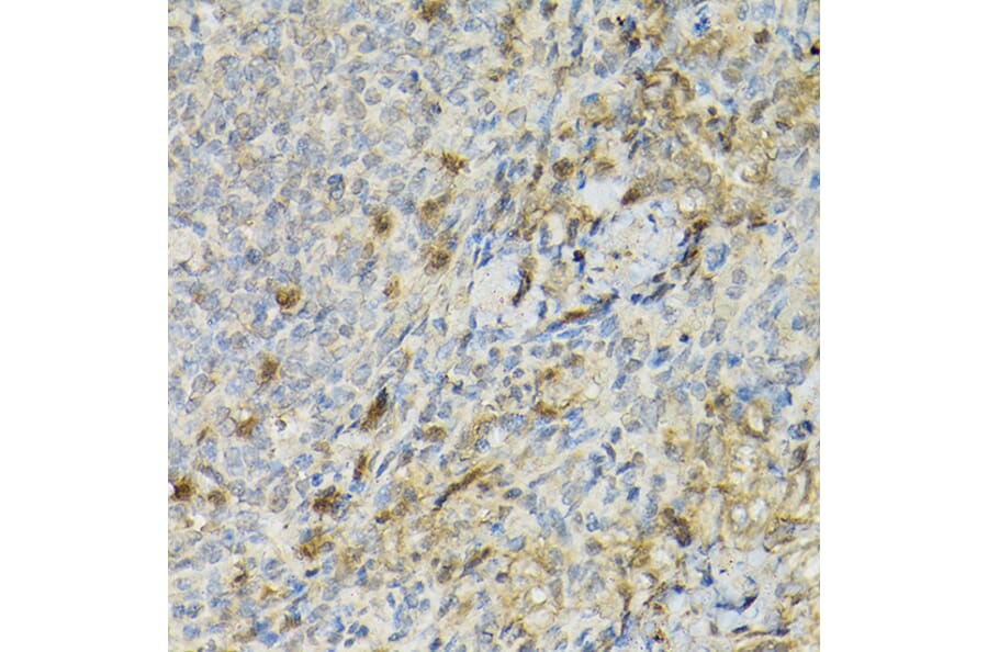
Immunohistochemistry analysis of paraffin-embedded rat spleen using Anti-IL-2 Receptor alpha Antibody (A14290) at a dilution of 1:100 (40x lens). Perform high pressure antigen retrieval with 10 mM citrate buffer pH 6.0 before commencing with IHC staining protocol.
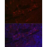
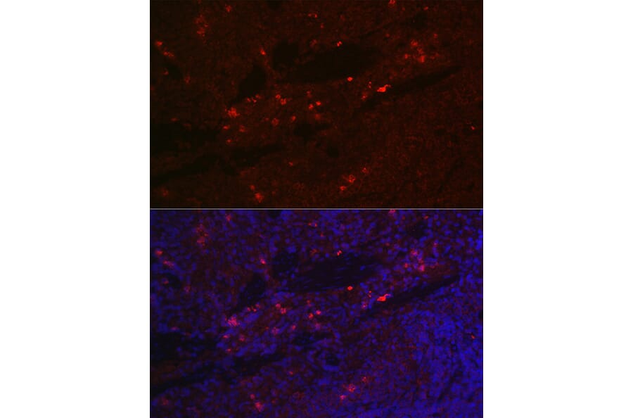
Immunofluorescence analysis of rat spleen cells using Anti-IL-2 Receptor alpha Antibody (A14290) at a dilution of 1:100 (40x lens). DAPI was used to stain the cell nuclei (blue).
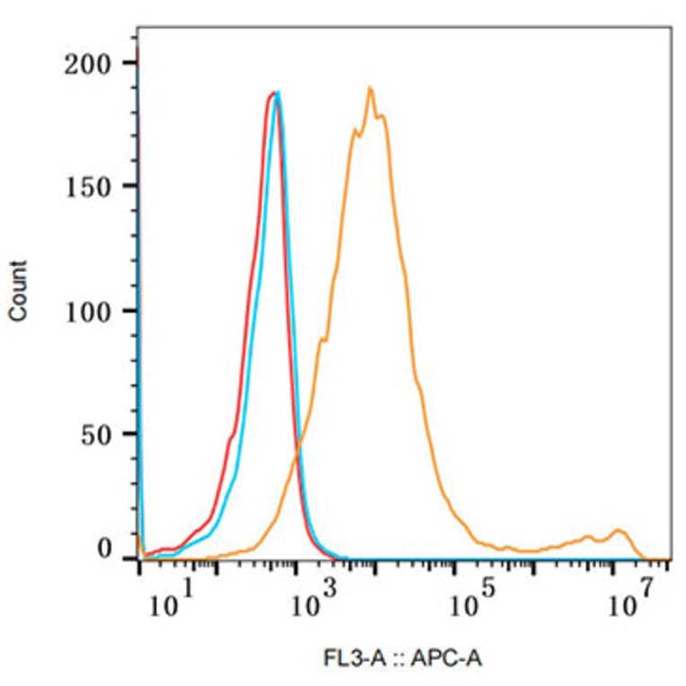
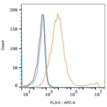
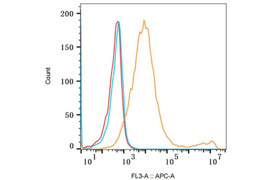
Flow cytometry analysis of 293F Transfection cells(+) and 293F cells(-), stained with Rabbit IgG isotype control (10 µg/ml, blue line) or Anti-IL-2 Receptor alpha Antibody (A14290), (2.5 µg/ml orange line), followed by Goat anti-Rabbit polyclonal antibody Alexa Fluor 647 (1:600 dilution) staining. Non-fluorescently stained Daudi cells was used as blank control (red line).