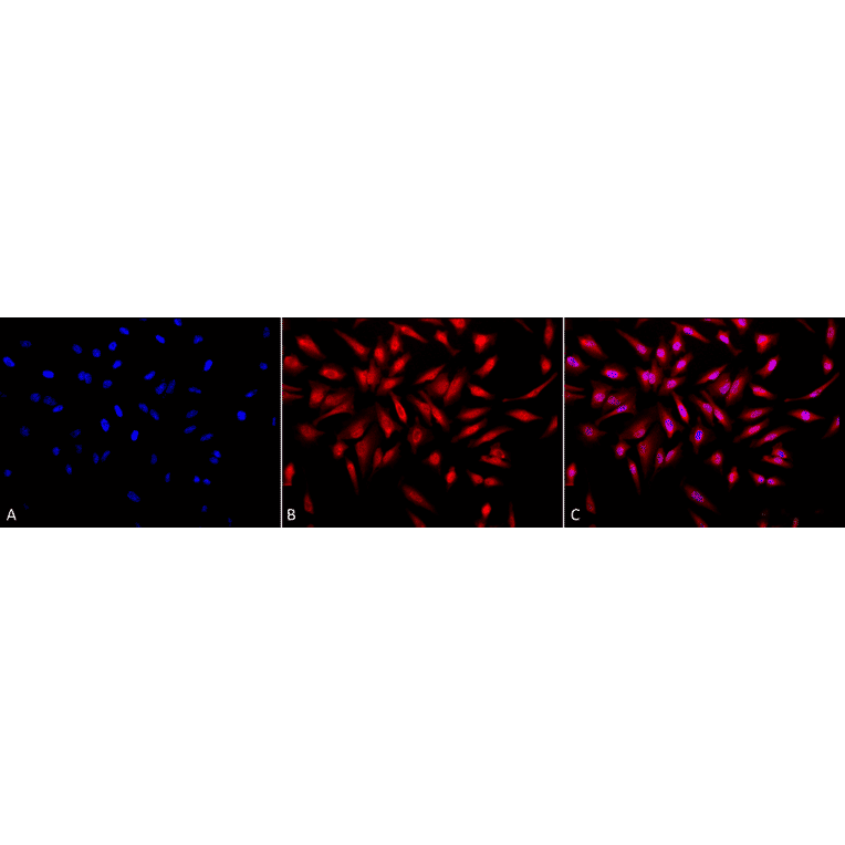
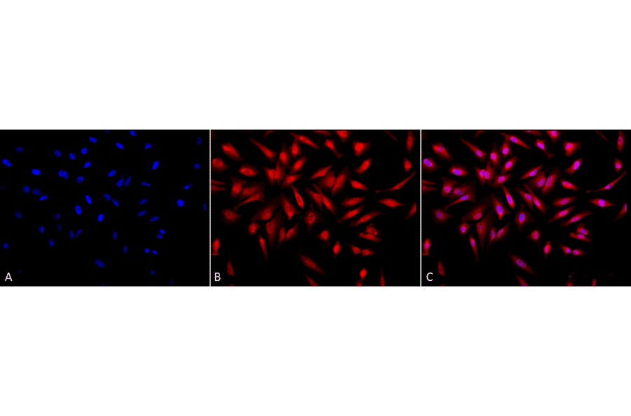
Immunocytochemistry/Immunofluorescence analysis of human heat shocked cervical cancer cell line (HeLa), fixed in 2% formaldehyde for 20 minutes at room temperature, using Anti-ING1 Antibody (A304909), at 1:50 for 12 hours at 4°C. The secondary antibody used was APC Goat Anti-Rabbit (red) at 1:200 for 2 hours at room temperature. Counterstain: DAPI (blue) nuclear stain at 1:40000 for 2 hours at room temperature. Localization: Nucleus. Cytoplasm. Magnification: 20x.(A) DAPI (blue) nuclear stain. (B) Anti-p33 ING1 Antibody. (C) Composite. Heat Shocked at 42°C for 30 min.
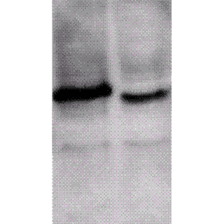
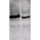
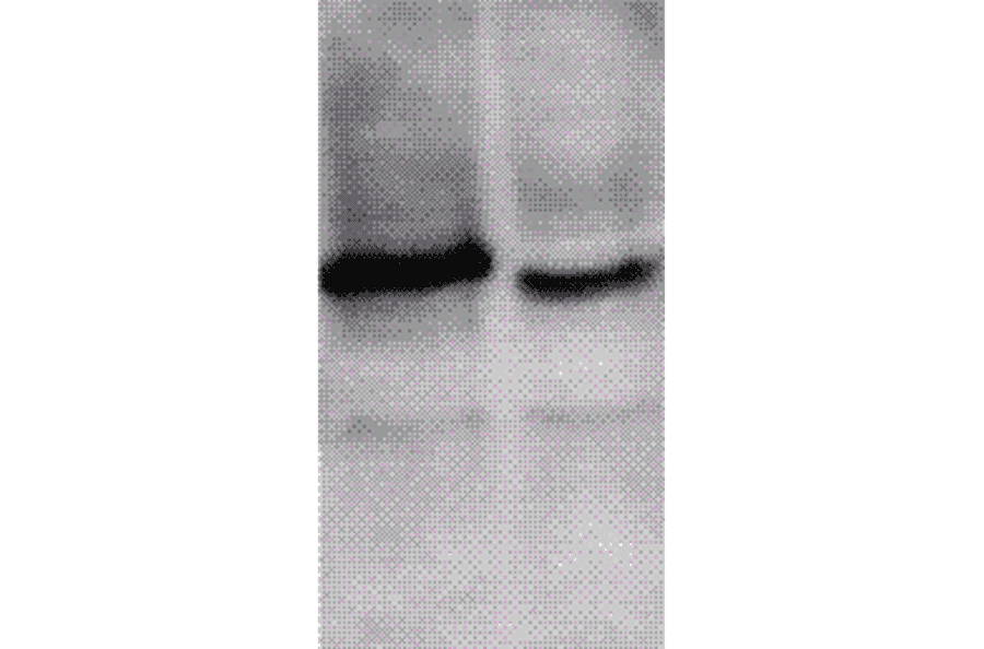
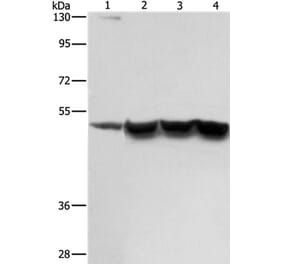
Western blot analysis of mouse brain cell lysates showing detection of ING1 protein using Anti-ING1 Antibody (A304909) at 1:1,000. Left: cell expressed flag-tag. Right: Untagged.
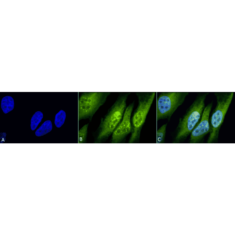
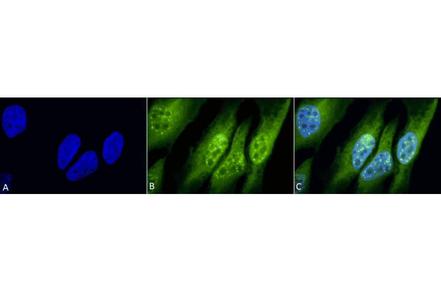
Immunocytochemistry/Immunofluorescence analysis of human heat shocked cervical cancer cell line (HeLa), fixed in 2% formaldehyde for 20 minutes at room temperature, using Anti-ING1 Antibody (A304909), at 1:50 for 12 hours at 4°C. The secondary antibody used was FITC Goat Anti-Rabbit (green) at 1:200 for 2 hours at room temperature. Counterstain: DAPI (blue) nuclear stain at 1:40000 for 2 hours at room temperature. Localization: Nucleus. Cytoplasm. Magnification: 100x.(A) DAPI (blue) nuclear stain. (B) Anti-p33 ING1 Antibody. (C) Composite. Heat Shocked at 42°C for 30 min.
Alternative products to Anti-ING1 Antibody (A304909)