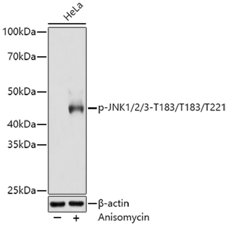
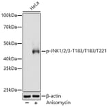
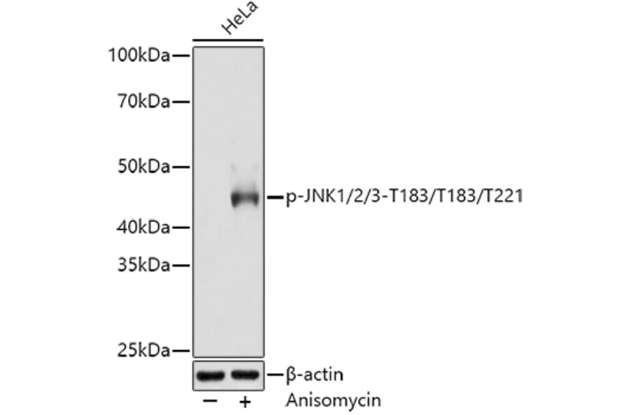
Western Blot - Anti-JNK1 + JNK2 + JNK3 (phospho Thr183 + Thr183 + Thr221) Antibody [ARC0193] (A80452)
Western blot analysis of extracts of HeLa cells, using Anti-JNK1 + JNK2 + JNK3 (phospho Thr183 + Thr183 + Thr221) Antibody (A80452) at 1:3,000 dilution. HeLa cells were treated by Anisomycin (25 ug/ml) at 37°C for 30 minutes after serum-starvation overnight. The secondary antibody was Goat Anti-Rabbit IgG H&L Antibody (HRP) at 1:10,000 dilution. Lysates/proteins were present at 25µg per lane. The blocking buffer used was 3% non-fat dry milk in TBST. Detection was with a ECL Basic Kit. Exposure time: 180s.
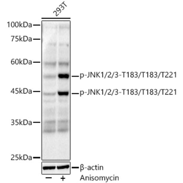
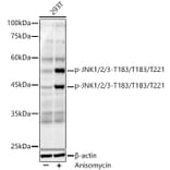
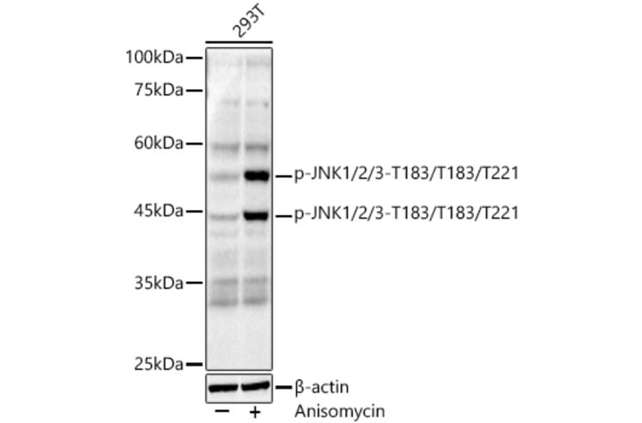
Western Blot - Anti-JNK1 + JNK2 + JNK3 (phospho Thr183 + Thr183 + Thr221) Antibody [ARC0193] (A80452)
Western blot analysis of extracts of 293T, using Anti-JNK1 + JNK2 + JNK3 (phospho Thr183 + Thr183 + Thr221) Antibody (A80452) at 1:1,000 dilution. 293T cells were treated by Anisomycin (25 ug/ml) at 37°C for 30 minutes after serum-starvation overnight. The secondary antibody was Goat Anti-Rabbit IgG H&L Antibody (HRP) at 1:10,000 dilution. Lysates/proteins were present at 25µg per lane. The blocking buffer used was 3% non-fat dry milk in TBST. Detection was with a ECL Basic Kit. Exposure time: 90s.
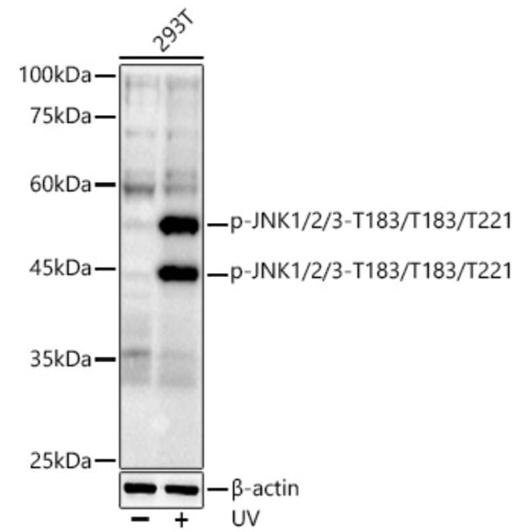
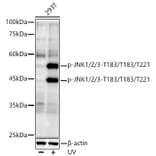
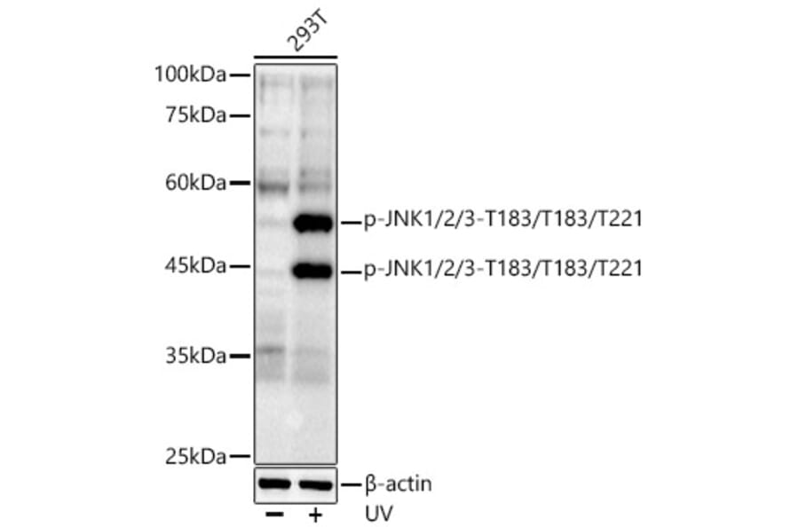
Western Blot - Anti-JNK1 + JNK2 + JNK3 (phospho Thr183 + Thr183 + Thr221) Antibody [ARC0193] (A80452)
Western blot analysis of extracts of 293T, using Anti-JNK1 + JNK2 + JNK3 (phospho Thr183 + Thr183 + Thr221) Antibody (A80452) at 1:1,000 dilution. 293T cells were treated by UV at room temperature for 15-30 minutes. The secondary antibody was Goat Anti-Rabbit IgG H&L Antibody (HRP) at 1:10,000 dilution. Lysates/proteins were present at 25µg per lane. The blocking buffer used was 3% non-fat dry milk in TBST. Detection was with a ECL Basic Kit. Exposure time: 90s.
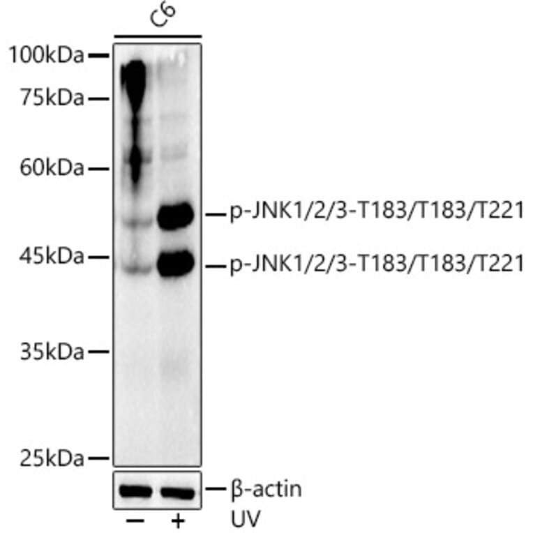
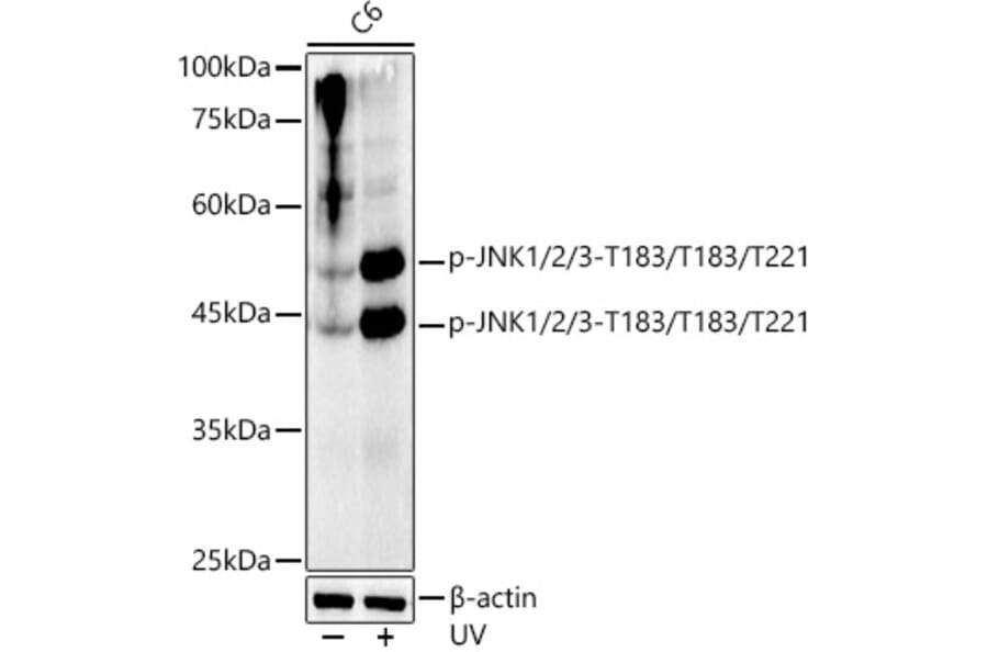
Western Blot - Anti-JNK1 + JNK2 + JNK3 (phospho Thr183 + Thr183 + Thr221) Antibody [ARC0193] (A80452)
Western blot analysis of extracts of C6, using Anti-JNK1 + JNK2 + JNK3 (phospho Thr183 + Thr183 + Thr221) Antibody (A80452) at 1:1,000 dilution. C6 cells were treated by UV at room temperature for 15-30 minutes. The secondary antibody was Goat Anti-Rabbit IgG H&L Antibody (HRP) at 1:10,000 dilution. Lysates/proteins were present at 25µg per lane. The blocking buffer used was 3% non-fat dry milk in TBST. Detection was with a ECL Basic Kit. Exposure time: 90s.
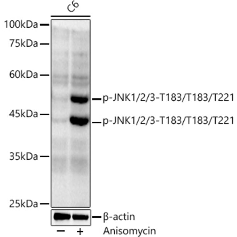
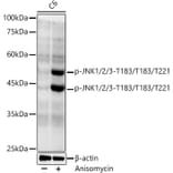
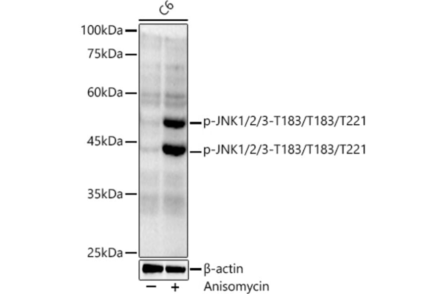
Western Blot - Anti-JNK1 + JNK2 + JNK3 (phospho Thr183 + Thr183 + Thr221) Antibody [ARC0193] (A80452)
Western blot analysis of extracts of C6, using Anti-JNK1 + JNK2 + JNK3 (phospho Thr183 + Thr183 + Thr221) Antibody (A80452) at 1:1,000 dilution. C6 cells were treated by Anisomycin (25 ug/ml) at 37°C for 20 minutes. The secondary antibody was Goat Anti-Rabbit IgG H&L Antibody (HRP) at 1:10,000 dilution. Lysates/proteins were present at 25µg per lane. The blocking buffer used was 3% non-fat dry milk in TBST. Detection was with a ECL Basic Kit. Exposure time: 180s.
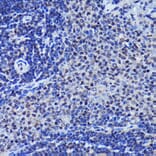
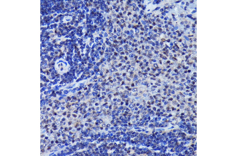
Immunohistochemistry analysis of paraffin-embedded rat spleen using Anti-JNK1 + JNK2 + JNK3 (phospho Thr183 + Thr183 + Thr221) Antibody (A80452) at a dilution of 1:100 (40x lens). Perform microwave antigen retrieval with 10 mM Tris/EDTA buffer pH 9.0 before commencing with IHC staining protocol.
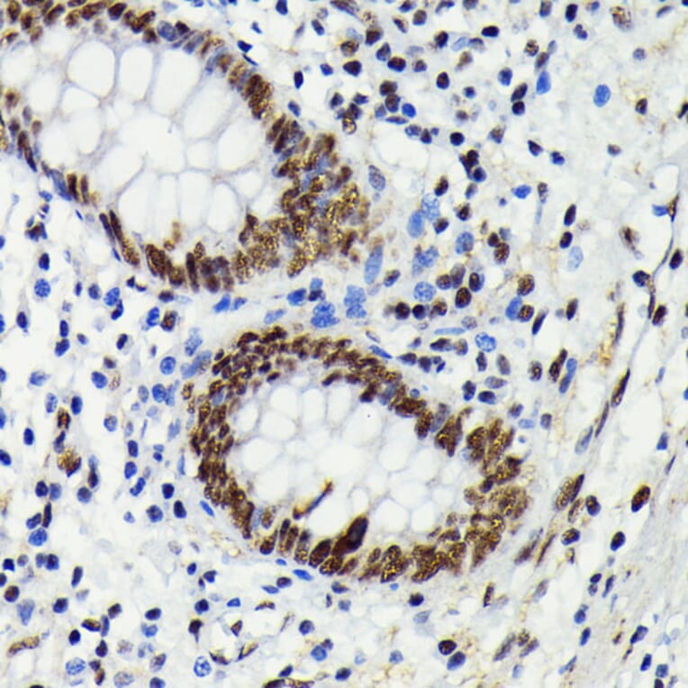
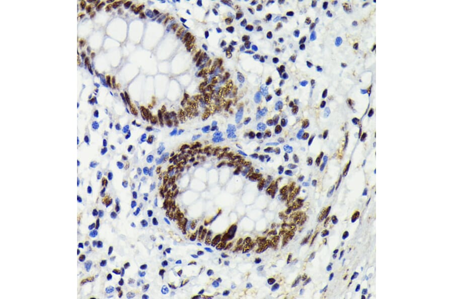
Immunohistochemistry analysis of paraffin-embedded human appendix tissue using Anti-JNK1 + JNK2 + JNK3 (phospho Thr183 + Thr183 + Thr221) Antibody (A80452) at a dilution of 1:100 (40x lens). Perform microwave antigen retrieval with 10 mM Tris/EDTA buffer pH 9.0 before commencing with IHC staining protocol.
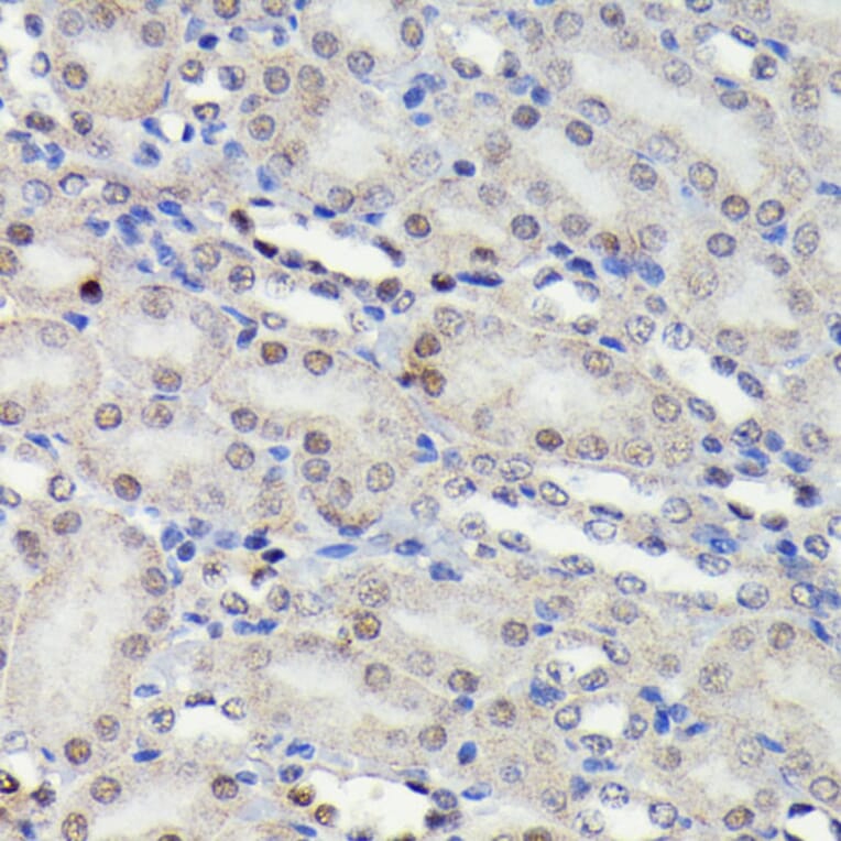
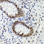
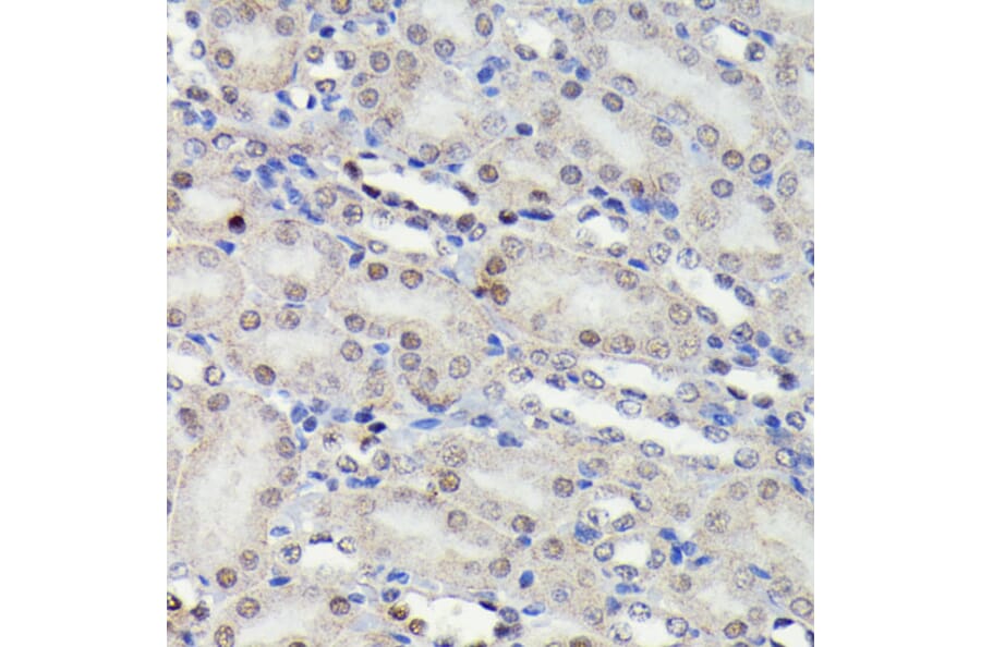
Immunohistochemistry analysis of paraffin-embedded mouse kidney using Anti-JNK1 + JNK2 + JNK3 (phospho Thr183 + Thr183 + Thr221) Antibody (A80452) at a dilution of 1:100 (40x lens). Perform microwave antigen retrieval with 10 mM Tris/EDTA buffer pH 9.0 before commencing with IHC staining protocol.
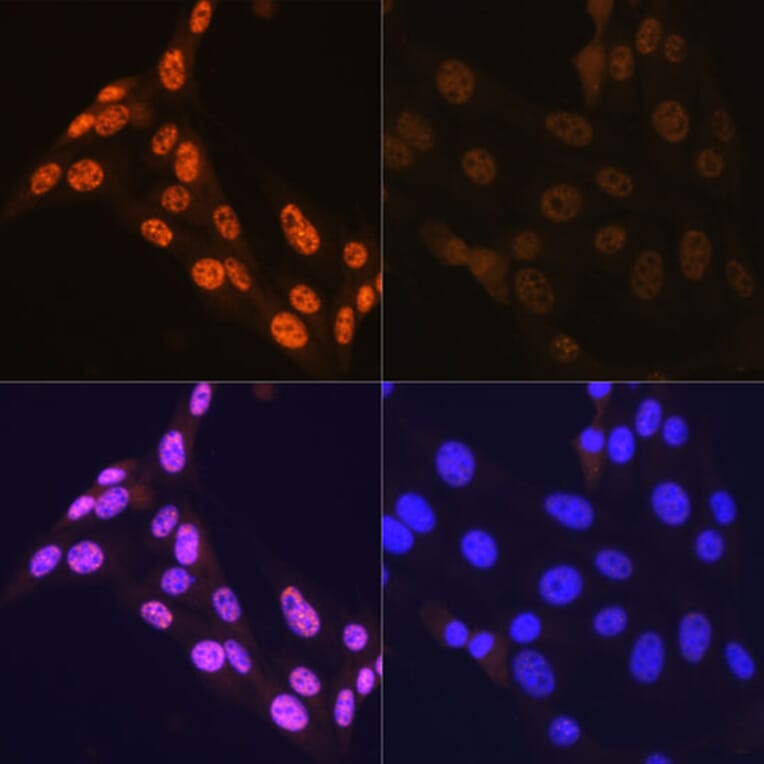
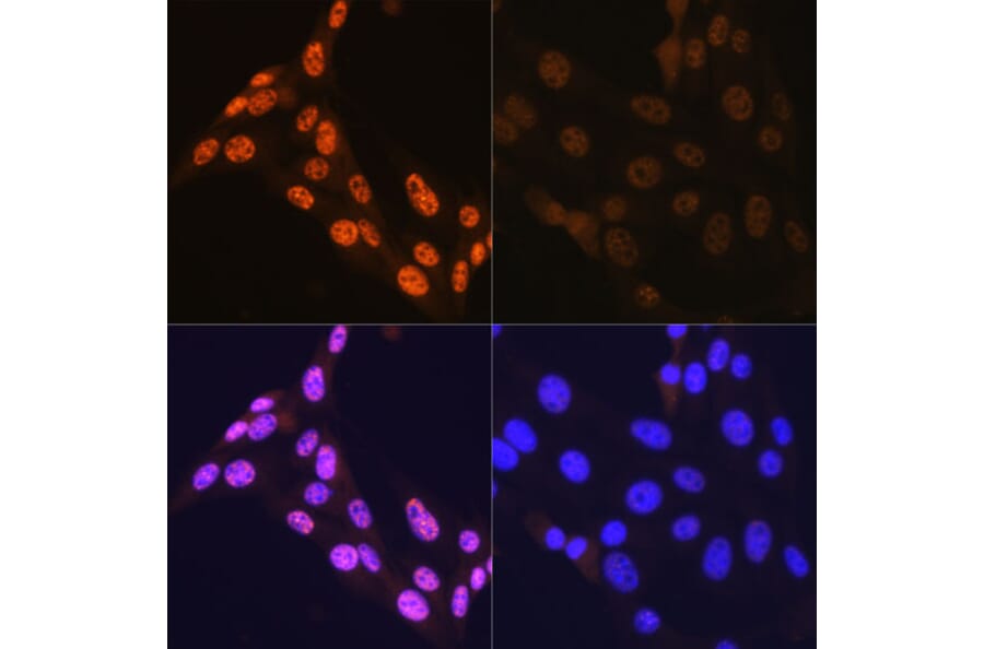
Immunofluorescence analysis of NIH-3T3 cells using Anti-JNK1 + JNK2 + JNK3 (phospho Thr183 + Thr183 + Thr221) Antibody (A80452). NIH-3T3 cells were treated by Anisomycin (25 ug/mL) at 37°C for 30 minutes after serum-starvation overnight. DAPI was used to stain the cell nuclei (blue).
Publishing research using Anti-JNK1 + JNK2 + JNK3 (phospho Thr183 + Thr183 + Thr221) Antibody [ARC0193] (A80452)? Please let us know so that we can list the citation on this page.