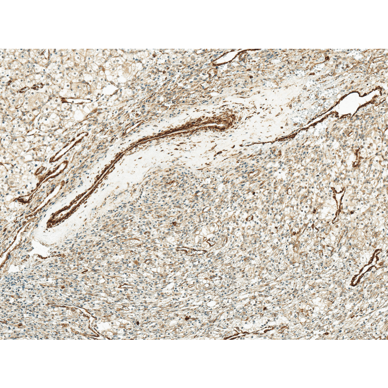
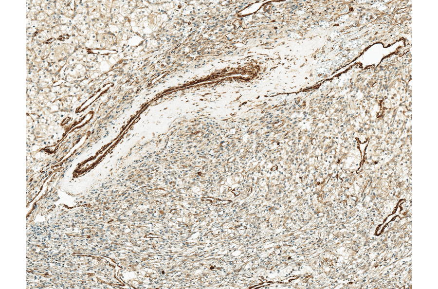
Immunohistochemistry analysis of human kidney, fixed in formalin and paraffin-embedded. The Primary Antibody used was Anti-Kir4.1 Antibody (A305164) at 1:50 for 30 minutes at room temperature. Counterstain: Hematoxylin. Magnification: 10X. HRP-DAB Detection.
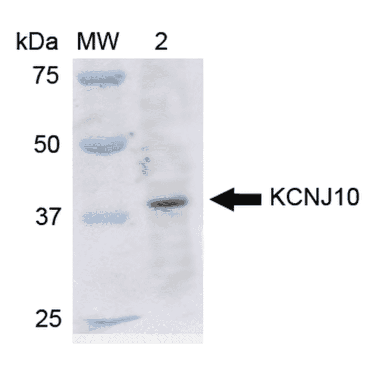
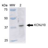
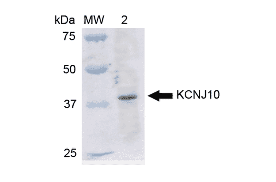
Western blot analysis of rat liver cell lysates showing detection of ~42.5 kDa Kir4.1 protein using Anti-Kir4.1 Antibody (A305164) at 1:1,000 for 2 hours at room temperature. Lane 1: Molecular Weight Ladder (MW). Lane 2: rat liver cell lysates. Load: 15 µg. Block: 5% Skim Milk in 1X TBST. The secondary antibody used was Goat Anti-Rabbit IgG: HRP at 1:2000 for 60 minutes at room temperature. Color Development: ECL solution for 6 min in room temperature. Predicted/Observed Size: ~42.5 kDa.
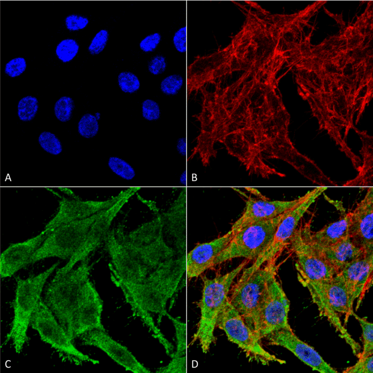
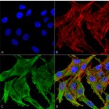
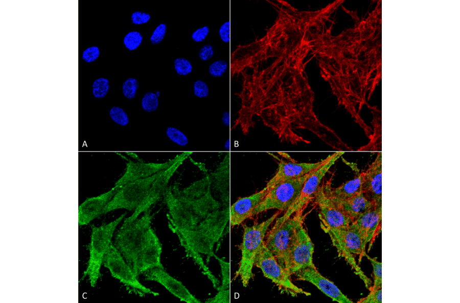
Immunocytochemistry/Immunofluorescence analysis of human colon carcinoma cell line (RKO), fixed in 4% formaldehyde for 15 min at room temperature, using Anti-Kir4.1 Antibody (A305164), at 1:100 for 60 minutes at room temperature. The secondary antibody used was Goat Anti-Rabbit ATTO 488 at 1:100 for 60 minutes at room temperature. Counterstain: Phalloidin Texas Red F-Actin stain; DAPI (blue) nuclear stain at 1:1000, 1:5,000 for 60 minutes at room temperature, 5 minutes at room temperature. Localization: Membrane, Cytoplasm. Magnification: 60X.(A) DAPI nuclear stain. (B) Phalloidin Texas Red F-Actin stain. (C) Kir4.1 Antibody. (D) Composite.
Publishing research using Anti-Kir4.1 Antibody (A305164)? Please let us know so that we can list the citation on this page.
Proteins predicted to interact with Kir4.1
Predicted protein interactions based upon String database. Revelancy score correlates with probability of interaction.