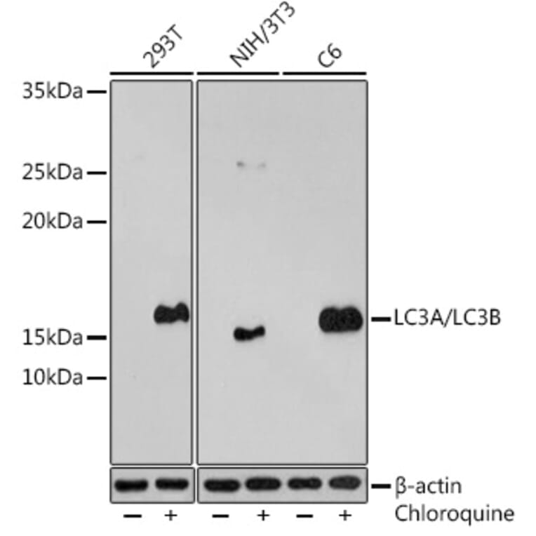
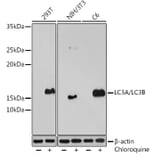
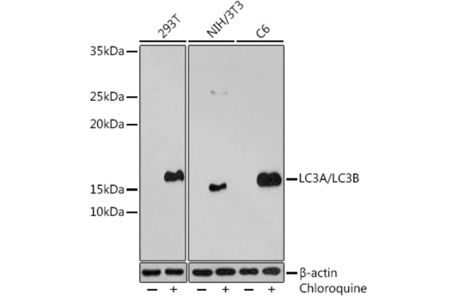
Western Blot - Anti-MAP1LC3A + LC3B Antibody (A14863)
Western blot analysis of extracts of various cell lines, using Anti-MAP1LC3A + LC3B Antibody (A14863) at 1:500 dilution. 293T cells were treated by Chloroquine (50 µM) at 37°C for 20 hours. NIH/3T3 cells were treated by Chloroquine (50 µM) at 37°C for 20 hours. C6 cells were treated by Chloroquine (50 µM) at 37°C for 20 hours. The secondary antibody was Goat Anti-Rabbit IgG H&L Antibody (HRP) at 1:10,000 dilution. Lysates/proteins were present at 25µg per lane. The blocking buffer used was 3% BSA. Detection was with a ECL Enhanced Kit. Exposure time: 3min.
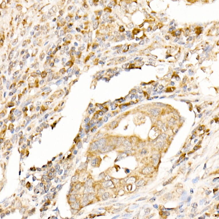
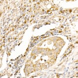
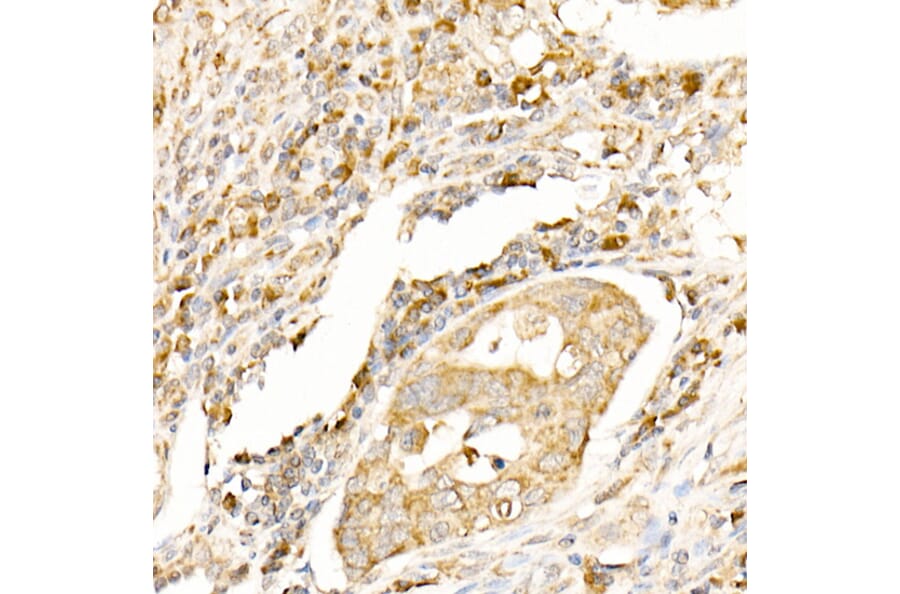
Immunohistochemistry analysis of paraffin-embedded human colon carcinoma tissue using Anti-MAP1LC3A + LC3B Antibody (A14863) at a dilution of 1:100 (40x lens). Perform high pressure antigen retrieval with 10 mM citrate buffer pH 6.0 before commencing with IHC staining protocol.
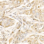
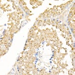
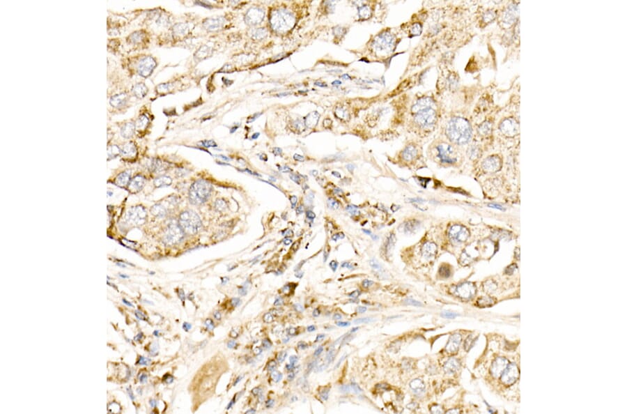
Immunohistochemistry analysis of paraffin-embedded human lung cancer using Anti-MAP1LC3A + LC3B Antibody (A14863) at a dilution of 1:100 (40x lens). Perform high pressure antigen retrieval with 10 mM citrate buffer pH 6.0 before commencing with IHC staining protocol.
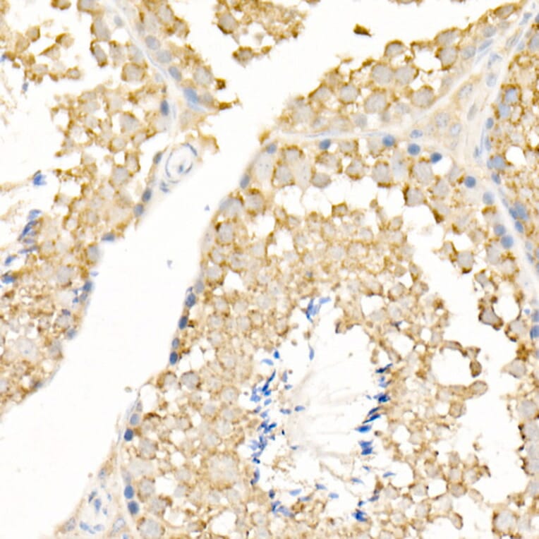
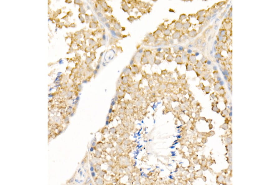
Immunohistochemistry analysis of paraffin-embedded mouse testis using Anti-MAP1LC3A + LC3B Antibody (A14863) at a dilution of 1:100 (40x lens). Perform high pressure antigen retrieval with 10 mM citrate buffer pH 6.0 before commencing with IHC staining protocol.
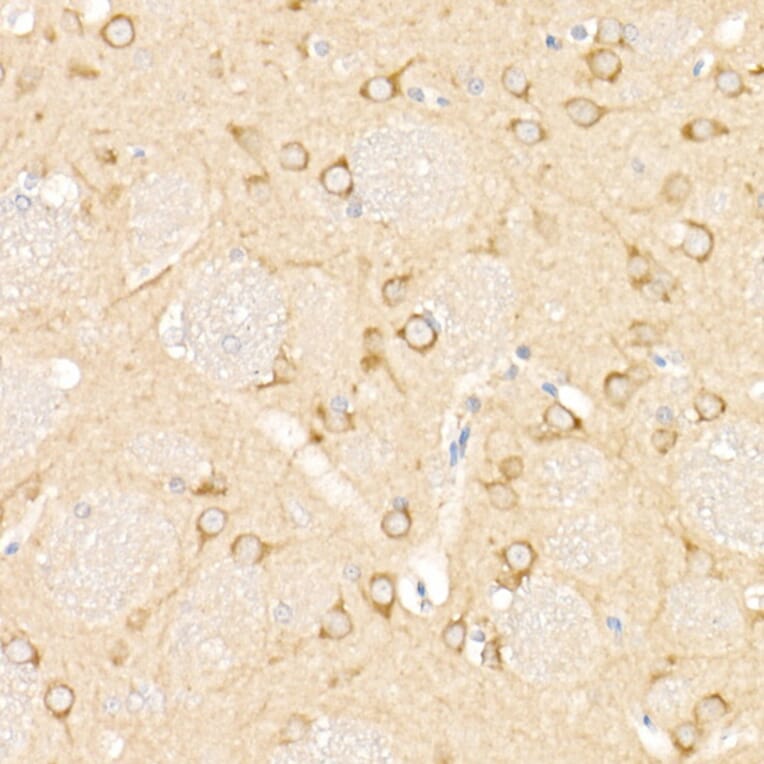
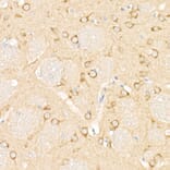
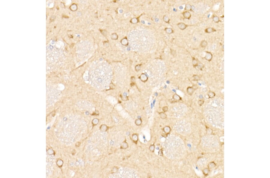
Immunohistochemistry analysis of paraffin-embedded rat brain using Anti-MAP1LC3A + LC3B Antibody (A14863) at a dilution of 1:100 (40x lens). Perform high pressure antigen retrieval with 10 mM citrate buffer pH 6.0 before commencing with IHC staining protocol.
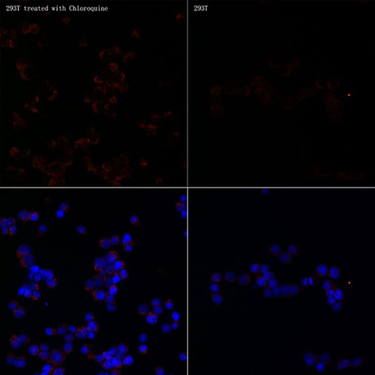
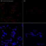
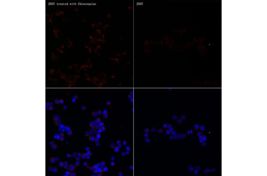
Immunofluorescence analysis of 293T Treated with Chloroquine and 293T cells using Anti-MAP1LC3A + LC3B Antibody (A14863) at a dilution of 1:100 (40x lens). DAPI was used to stain the cell nuclei (blue).