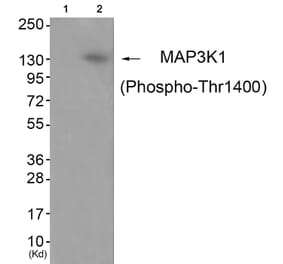
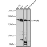
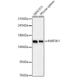
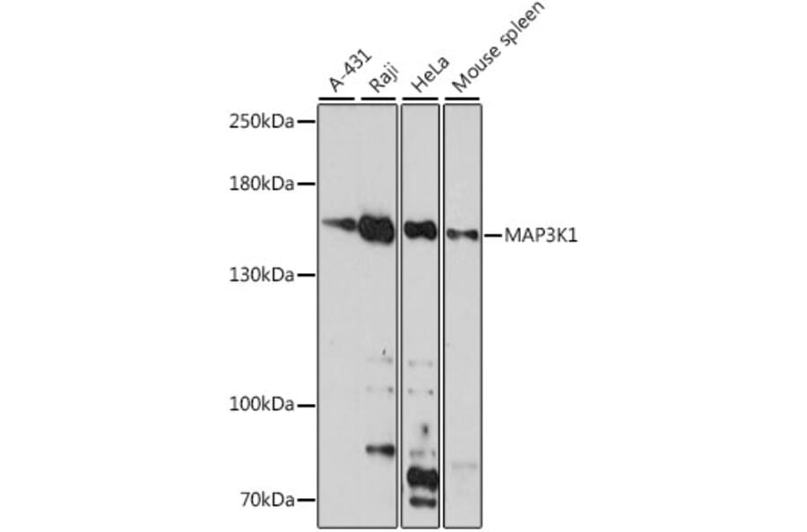
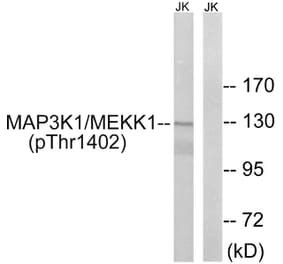
MAP3K1 expression in various lysates analyzed by western blot. Primary antibody incubation was performed for 1 hour on 25ug protein per lane with Anti-MAP3K1 Antibody (A88425) at a dilution of 1:1000 and detected with chemiluminescence.
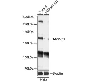
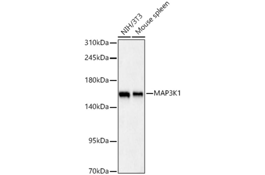
MAP3K1 expression in various lysates analyzed by western blot. Primary antibody incubation was performed for 1 hour on 25ug protein per lane with Anti-MAP3K1 Antibody (A88425) at a dilution of 1:1000 and detected with chemiluminescence.
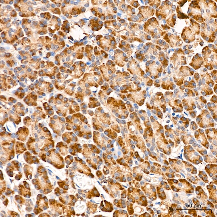
MAP3K1 expression in human pancreas tissue analyzed by immunohistochemistry. Tissue was paraffin-embedded, and antigen retrieval was achieved with 10 mM citrate buffer, pH 6.0, under high pressure. Staining was performed with Anti-MAP3K1 Antibody (A88425) at a dilution of 1:300.
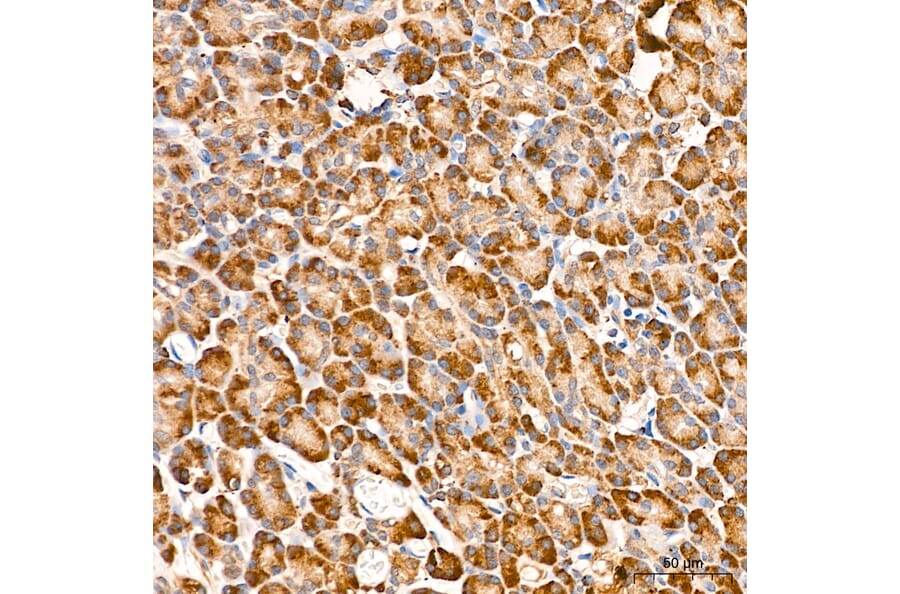
MAP3K1 expression in human liver tissue analyzed by immunohistochemistry. Tissue was paraffin-embedded, and antigen retrieval was achieved with 10 mM citrate buffer, pH 6.0, under high pressure. Staining was performed with Anti-MAP3K1 Antibody (A88425) at a dilution of 1:300.
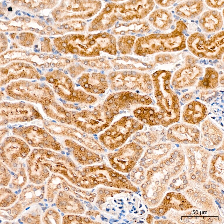
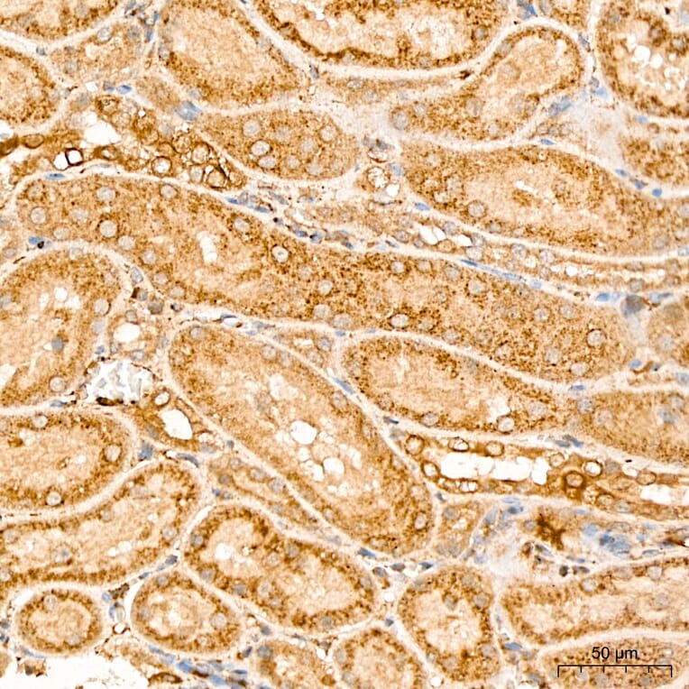
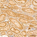
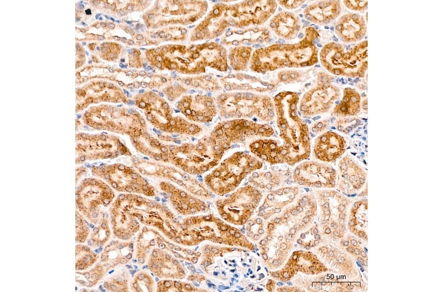
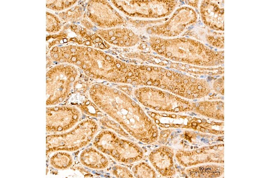
MAP3K1 expression in mouse kidney tissue analyzed by immunohistochemistry. Tissue was paraffin-embedded, and antigen retrieval was achieved with 10 mM citrate buffer, pH 6.0, under high pressure. Staining was performed with Anti-MAP3K1 Antibody (A88425) at a dilution of 1:300.
MAP3K1 expression in rat kidney tissue analyzed by immunohistochemistry. Tissue was paraffin-embedded, and antigen retrieval was achieved with 10 mM citrate buffer, pH 6.0, under high pressure. Staining was performed with Anti-MAP3K1 Antibody (A88425) at a dilution of 1:300.
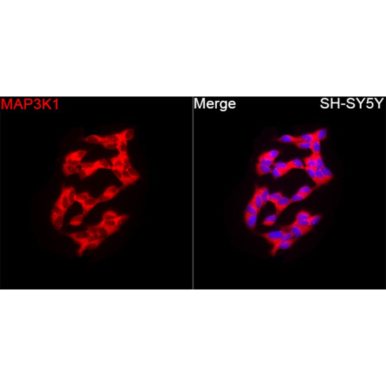
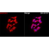
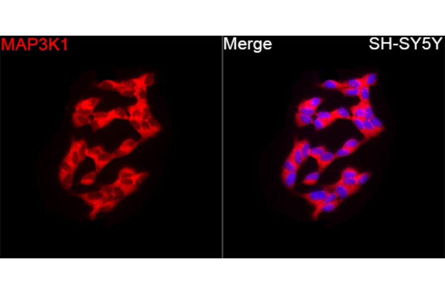
MAP3K1 expression in SH-SY5Y cells analyzed by immunofluorescence. Staining was performed with Anti-MAP3K1 Antibody (A88425) at a dilution of 1:200 followed by Cy3-conjugated Goat anti-Rabbit IgG (H+L)secondary antibody at a dilution of 1:500. Nuclei were stained with DAPI (blue).
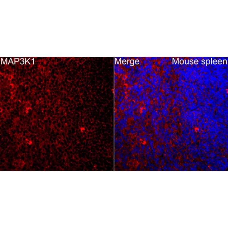
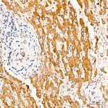
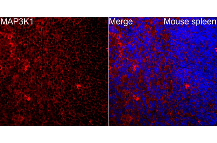
MAP3K1 expression in Mouse spleen tissue analyzed by immunofluorescence. Staining was performed with Anti-MAP3K1 Antibody (A88425) at a dilution of 1:200 followed by Cy3-conjugated Goat anti-Rabbit IgG (H+L)secondary antibody at a dilution of 1:500. Nuclei were stained with DAPI (blue).


![Immunohistochemistry - Anti-MAP3K1 Antibody [2F6] (A251815) - Antibodies.com](https://cdn.antibodies.com/image/catalog/249/A249314_1.jpg?profile=product_alternative)
![Immunohistochemistry - Anti-MAP3K1 Antibody [2F6] - BSA and Azide free (A249314) - Antibodies.com](https://cdn.antibodies.com/image/catalog/252/A252494_1.jpg?profile=product_alternative)