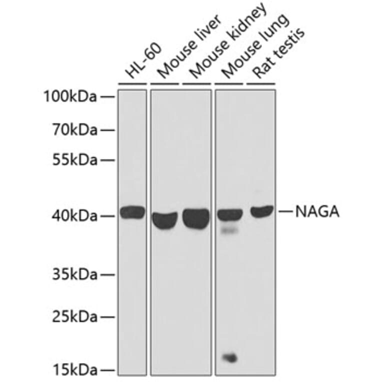
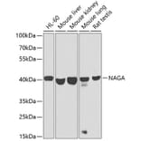
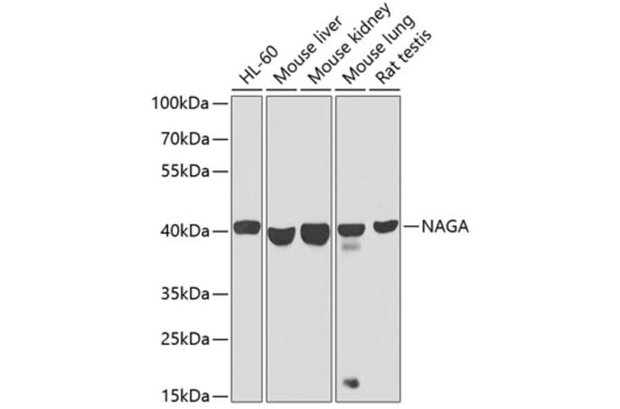
Western blot analysis of extracts of various cell lines, using Anti-NAGA Antibody (A16379) at 1:1,000 dilution. The secondary antibody was Goat Anti-Rabbit IgG H&L Antibody (HRP) at 1:10,000 dilution. Lysates/proteins were present at 25µg per lane. The blocking buffer used was 3% non-fat dry milk in TBST. Detection was with a ECL Basic Kit. Exposure time: 30s.
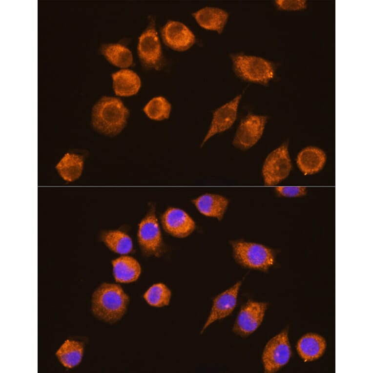
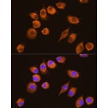
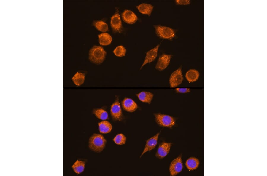
Immunofluorescence analysis of L929 cells using Anti-NAGA Antibody (A16379) at a dilution of 1:100 (40x lens). DAPI was used to stain the cell nuclei (blue).
Publishing research using Anti-NAGA Antibody (A16379)? Please let us know so that we can list the citation on this page.
Proteins predicted to interact with NAGA
Predicted protein interactions based upon String database. Revelancy score correlates with probability of interaction.