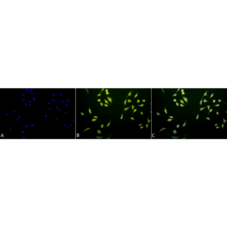
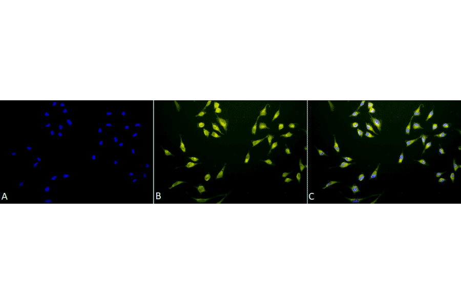
Immunocytochemistry/Immunofluorescence analysis of human cervical cancer cell line (HeLa), fixed in 2% formaldehyde for 20 minutes at room temperature, using Anti-P70 S6 Kinase beta / SRK Antibody (A305084), at 1:50 for 12 hours at 4°C. The secondary antibody used was R-PE Goat Anti-Rabbit (yellow) at 1:200 for 2 hours at room temperature. Counterstain: DAPI (blue) nuclear stain at 1:40000 for 2 hours at room temperature. Localization: Cytoplasm. Perinuclear region of cytoplasm. Magnification: 20x.(A) DAPI (blue) nuclear stain. (B) Anti-p70 S6K Antibody. (C) Composite.
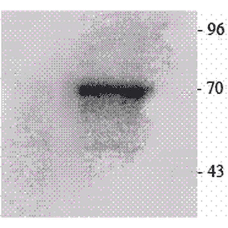
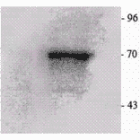
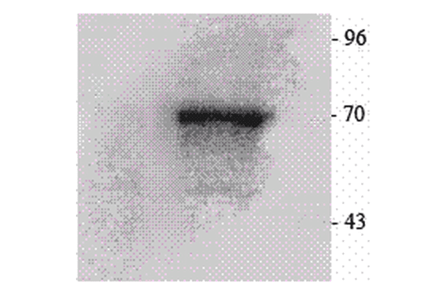
Western Blot - Anti-P70 S6 Kinase beta / SRK Antibody (A305084)
Western blot analysis of mouse brain cell lysates showing detection of p70 S6K protein using Anti-P70 S6 Kinase beta / SRK Antibody (A305084) at 1:1,000. Anti-p70 S6K Antibody on right, IP negative control on the left.
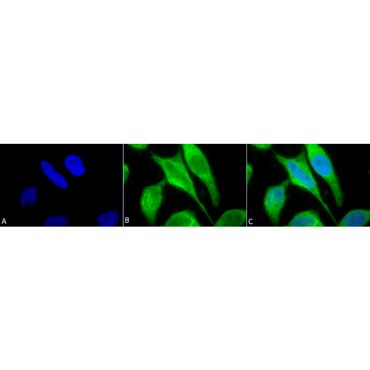
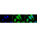
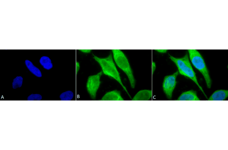
Immunocytochemistry/Immunofluorescence analysis of human cervical cancer cell line (HeLa), fixed in 2% formaldehyde for 20 minutes at room temperature, using Anti-P70 S6 Kinase beta / SRK Antibody (A305084), at 1:50 for 12 hours at 4°C. The secondary antibody used was FITC Goat Anti-Rabbit (green) at 1:200 for 2 hours at room temperature. Counterstain: DAPI (blue) nuclear stain at 1:40000 for 2 hours at room temperature. Localization: Cytoplasm. Perinuclear region of cytoplasm. Magnification: 100x.(A) DAPI (blue) nuclear stain. (B) Anti-p70 S6K Antibody. (C) Composite.