Supplied in Phosphate Buffered Saline, pH 7.3, with 50% Glycerol, 0.05% BSA, and 0.02% Sodium Azide.
Storage
Shipped at 4°C. Upon delivery aliquot and store at -20°C. Avoid freeze/thaw cycles.
Synonyms
Nuclear poly(A)-binding protein 1, PAB2, PABII, PABP-2, PABP2, Poly(A)-binding protein 2, Poly(A)-binding protein II, Polyadenylate-binding nuclear protein 1, Polyadenylate-binding protein 2
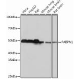
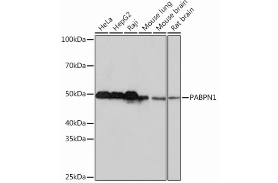
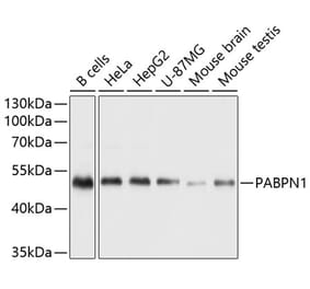
Western Blot - Anti-PABPN1 Antibody [ARC0730] (A306775)
Western blot analysis of extracts of various cell lines, using Anti-polyclonal antibodyPN1 Antibody (A306775) at 1:1,000 dilution. The secondary antibody was Goat Anti-Rabbit IgG H&L Antibody (HRP) at 1:10,000 dilution. Lysates/proteins were present at 25µg per lane. The blocking buffer used was 3% non-fat dry milk in TBST. Detection was with a ECL Basic Kit. Exposure time: 10s.
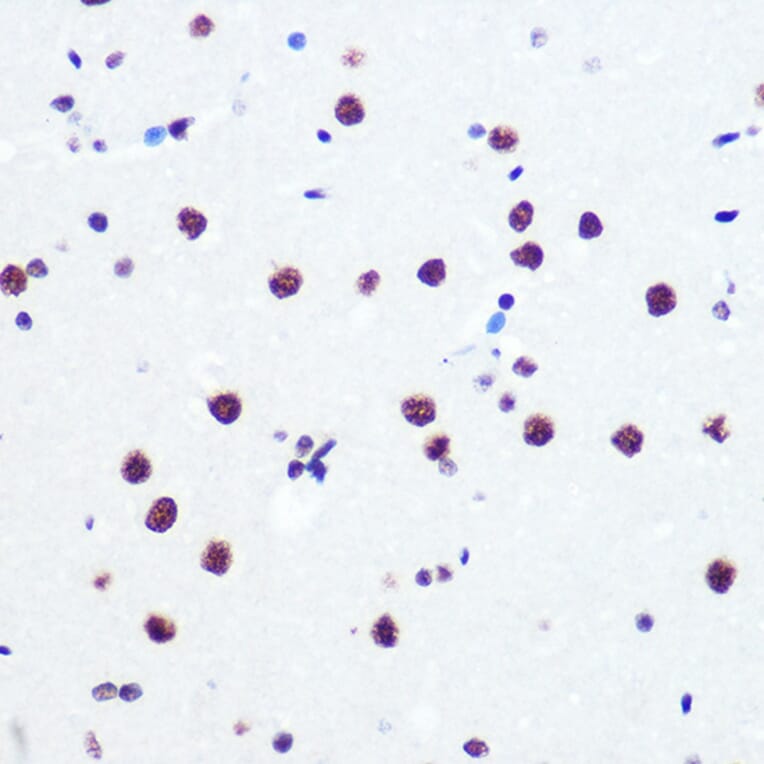
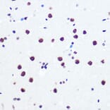
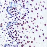
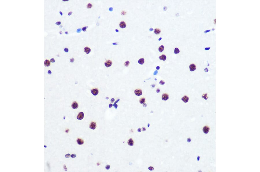
Immunohistochemistry analysis of paraffin-embedded rat brain using Anti-polyclonal antibodyPN1 Antibody (A306775) at a dilution of 1:100 (40x lens). Perform microwave antigen retrieval with 10 mM PBS buffer pH 7.2 before commencing with IHC staining protocol.
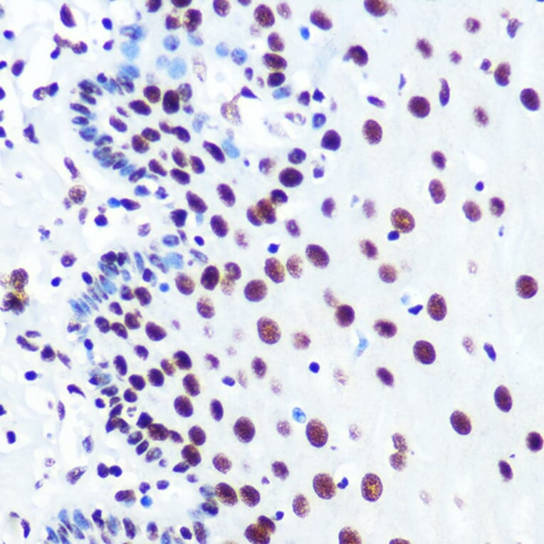
Immunohistochemistry analysis of paraffin-embedded human esophageal using Anti-polyclonal antibodyPN1 Antibody (A306775) at a dilution of 1:100 (40x lens). Perform microwave antigen retrieval with 10 mM PBS buffer pH 7.2 before commencing with IHC staining protocol.
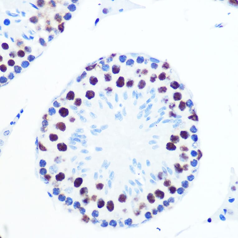
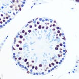
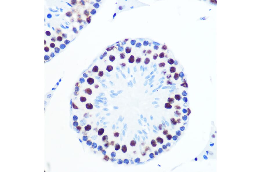
Immunohistochemistry analysis of paraffin-embedded mouse testis using Anti-polyclonal antibodyPN1 Antibody (A306775) at a dilution of 1:100 (40x lens). Perform microwave antigen retrieval with 10 mM PBS buffer pH 7.2 before commencing with IHC staining protocol.
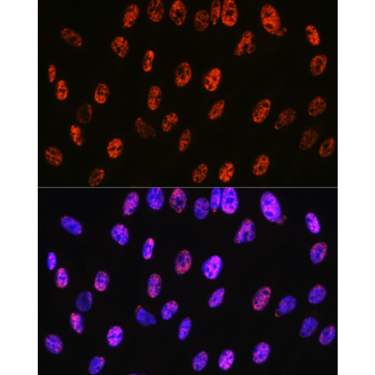
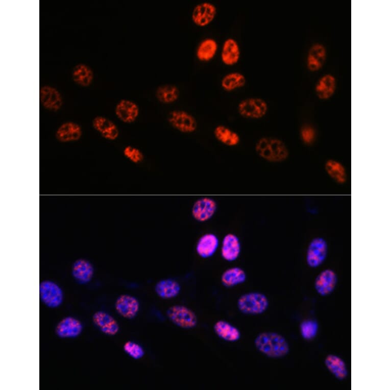
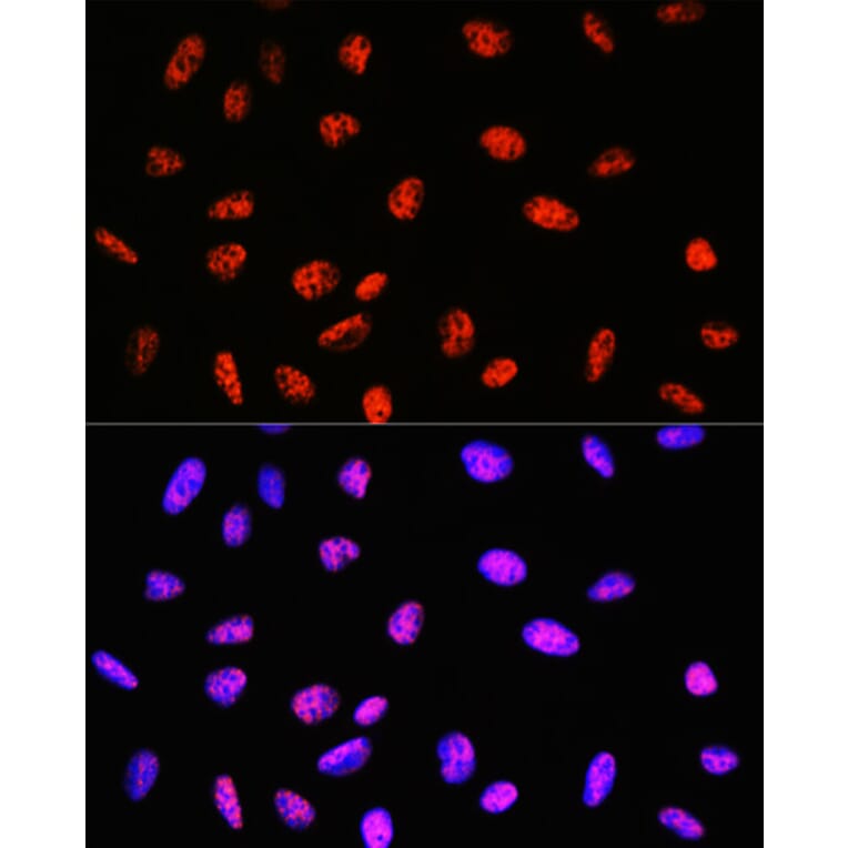
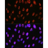
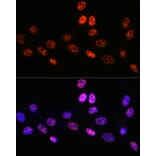
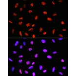
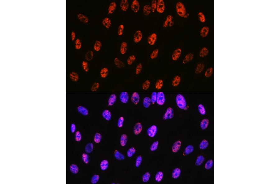
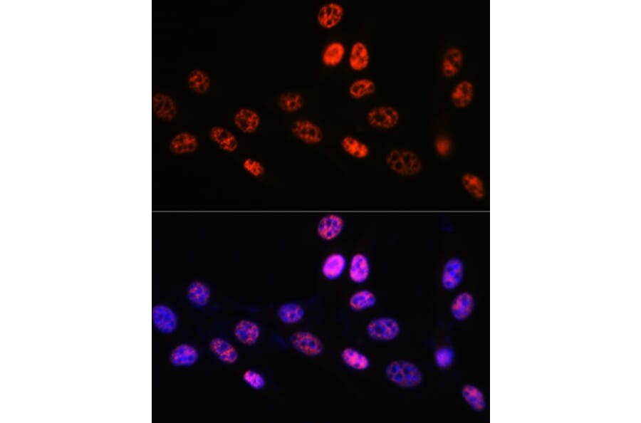
Immunofluorescence analysis of C6 cells using Anti-polyclonal antibodyPN1 Antibody (A306775) at a dilution of 1:100 (40x lens). DAPI was used to stain the cell nuclei (blue).
Immunofluorescence analysis of NIH-3T3 cells using Anti-polyclonal antibodyPN1 Antibody (A306775) at a dilution of 1:100 (40x lens). DAPI was used to stain the cell nuclei (blue).
Immunofluorescence analysis of U-2 OS cells using Anti-polyclonal antibodyPN1 Antibody (A306775) at a dilution of 1:100 (40x lens). DAPI was used to stain the cell nuclei (blue).