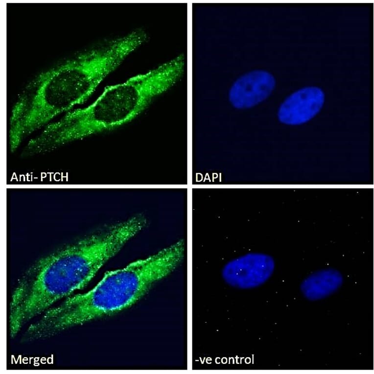
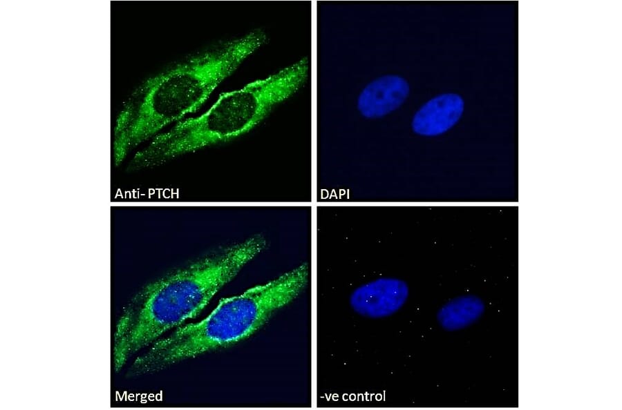
Patched/PTCH1 expression in HeLa cells analyzed by immunofluorescence. Cells were permeabilized with 0.15% Triton. Staining was performed with Anti-Patched/PTCH1 Antibody (A83443) at 10µg/ml for 1 hour and Alexa Fluor 488 secondary antibody at 2µg/ml. Cytoplasmic/Golgi apparatus staining shown and nuclei were stained with DAPI (blue). Negative control: Goat IgG Isotype Control at 10µg/ml followed by Alexa Fluor 488 secondary antibody at 2µg/ml.
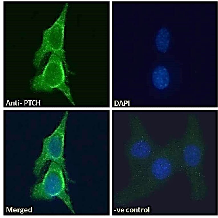
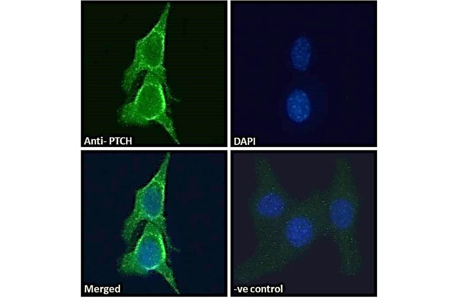
Patched/PTCH1 expression in NIH3T3 cells analyzed by immunofluorescence. Cells were permeabilized with 0.15% Triton. Staining was performed with Anti-Patched/PTCH1 Antibody (A83443) at 10µg/ml for 1 hour and Alexa Fluor 488 secondary antibody at 2µg/ml. Golgi apparatus staining shown and nuclei were stained with DAPI (blue). Negative control: Goat IgG Isotype Control at 10µg/ml followed by Alexa Fluor 488 secondary antibody at 2µg/ml.
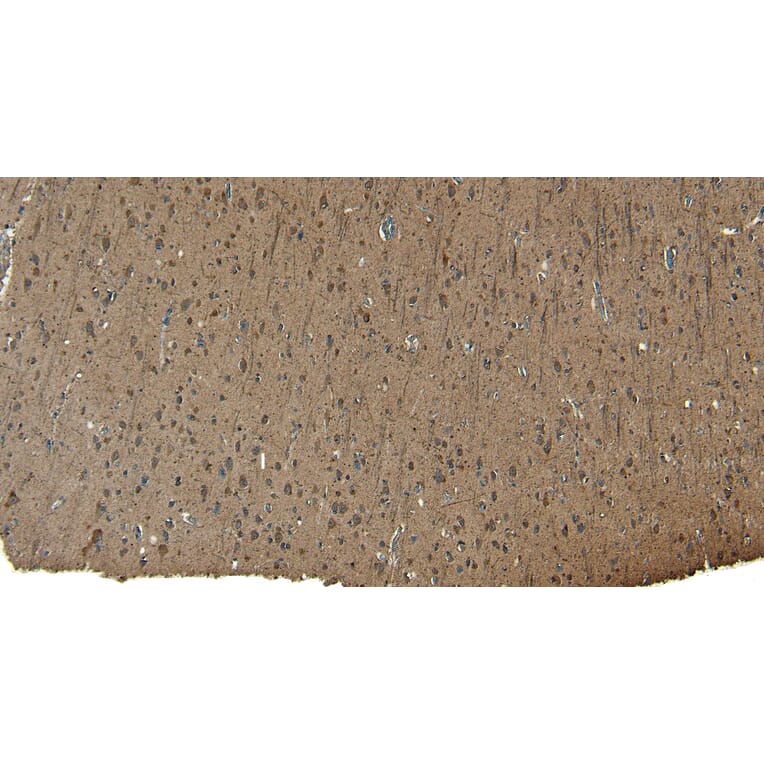
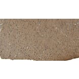
Patched/PTCH1 expression in Human Cortex analyzed by immunohistochemistry. Tissue was paraffin-embedded, and antigen retrieval was achieved by heating in citrate buffer, pH 6. Staining was performed with Anti-Patched/PTCH1 Antibody (A83443) at 8µg/ml and revealed with horseradish peroxidase (HRP).
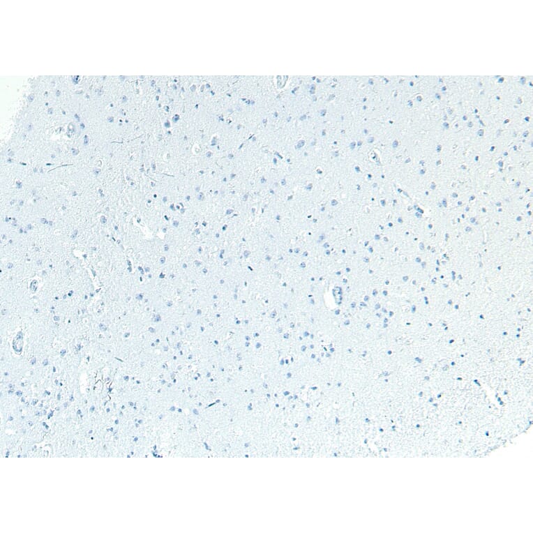
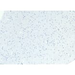
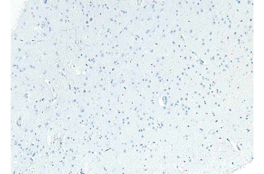
Negative control for Patched/PTCH1 expression in Human Cortex analyzed by immunohistochemistry. Tissue was paraffin-embedded, and staining procedure was performed in the absence of primary antibody.
Publishing research using Anti-Patched/PTCH1 Antibody (A83443)? Please let us know so that we can list the citation on this page.