
Unconjugated
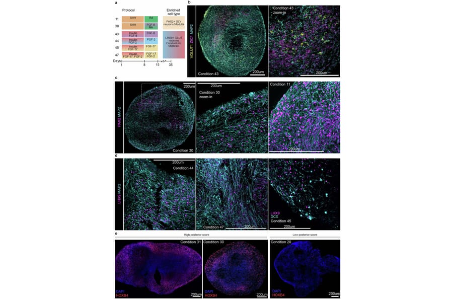
Patterning of the neural tube establishes midbrain and hindbrain structures that coordinate motor movement, process sensory input and integrate cognitive functions. Cellular impairment within these structures underlies diverse neurological disorders, and in vitro organoid models promise inroads to understanding development and modeling disease. Here, we use paired single-cell transcriptome and accessible chromatin sequencing to map cell composition and regulatory mechanisms in organoid models of midbrain and hindbrain. We find that existing midbrain organoid protocols generate ventral and dorsal cell types, covering regions including floor plate, dorsal and ventral midbrain and adjacent hindbrain regions. Gene regulatory network inference and transcription factor perturbation resolve mechanisms underlying neuronal differentiation. A single-cell multiplexed patterning screen identifies morphogen concentrations that expand existing organoid models, including conditions generating medulla glycinergic neurons and cerebellum glutamatergic subtypes. Together, the multi-omic atlas and morphogen screen reveal morphogen-regulon relationships guiding region-specific progenitor differentiation towards diverse neuron types of the posterior brain.
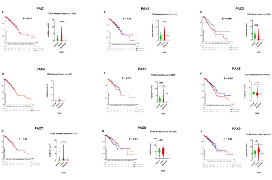
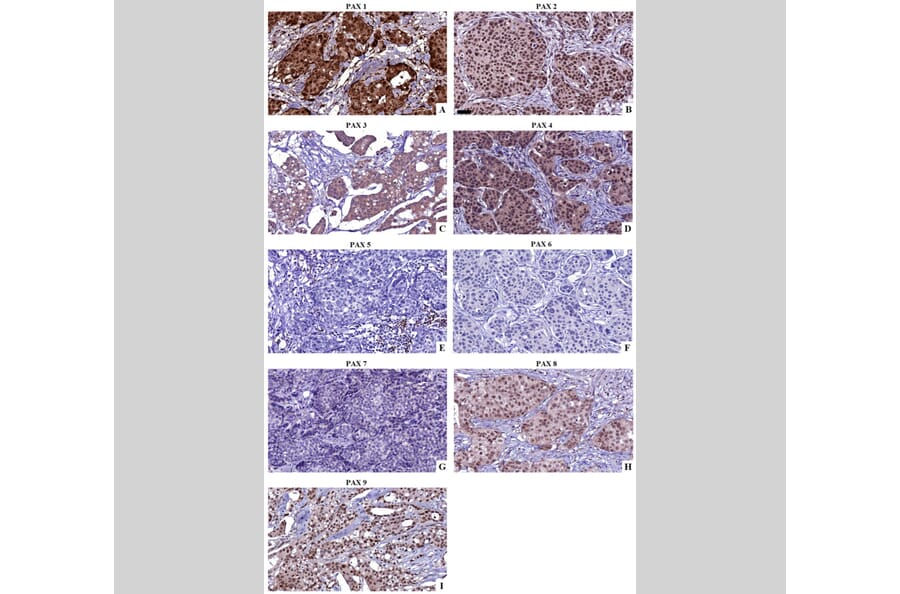
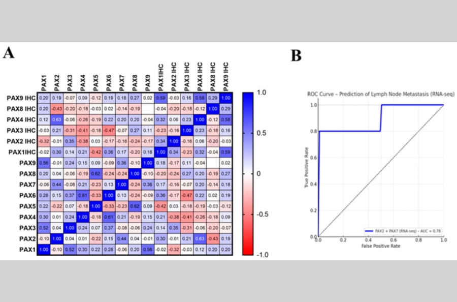
Breast cancer is the leading cause of cancer-related death in women, and despite advances in preventive screening as well as in molecular classification, many patients still do not benefit from existing therapies, highlighting the importance of identifying new molecular determinants of treatment resistance. The Paired-box (PAX) family of developmental transcription factors are central regulators of tissue morphogenesis and lineage specification, yet their reactivation in tumors and contribution to breast cancer progression remain only partially defined. Here, a multi-level analysis integrating RNA sequencing and protein profiling in twenty-one primary breast carcinomas shows that distinct PAX members are directly correlated to distinct fundamental cancer hallmarks, including proliferation, cell death, epithelial-mesenchymal transition, immune evasion, and genomic instability. Specifically, PAX1 and PAX9 correlates with both cell death and proliferative markers, indicating dual roles in the regulation of cell fate. PAX1 and PAX9 correlate with both proliferative and apoptotic markers, indicating dual roles in cell fate regulation. PAX3, PAX5, and PAX8 are mainly associated with immune checkpoint expression, including PD-L1 and TIGIT, while PAX6 is linked to microsatellite instability and tumor mutational burden, implicating it in genomic dysregulation. Therefore, PAX-based molecular signatures identify that accurately predict lymph node metastasis at transcriptomic (PAX2-PAX7) levels. These findings establish PAX transcription factors as key modulators of breast cancer biology and support their potential as clinically relevant biomarkers for prognostic refinement and therapeutic stratification.
![Immunohistochemistry - Anti-PAX2 Antibody [PAX2/1104] - BSA and Azide free (A249559) - Antibodies.com](https://cdn.antibodies.com/image/catalog/252/A252739_1.jpg?profile=product_top)
![Immunohistochemistry - Anti-PAX2 Antibody [PAX2/1104] - BSA and Azide free (A249559) - Antibodies.com](https://cdn.antibodies.com/image/catalog/252/A252739_2.jpg?profile=product_top)
![Immunohistochemistry - Anti-PAX2 Antibody [PAX2/1104] - BSA and Azide free (A249559) - Antibodies.com](https://cdn.antibodies.com/image/catalog/252/A252739_3.jpg?profile=product_top)
![SDS-PAGE - Anti-PAX2 Antibody [PAX2/1104] - BSA and Azide free (A249559) - Antibodies.com](https://cdn.antibodies.com/image/catalog/252/A252739_4.jpg?profile=product_top)
![Protein Array - Anti-PAX2 Antibody [PAX2/1104] - BSA and Azide free (A249559) - Antibodies.com](https://cdn.antibodies.com/image/catalog/252/A252739_5.jpg?profile=product_top)
![Immunofluorescence - Anti-PAX2 Antibody [PAX2/1104] - BSA and Azide free (A252739) - Antibodies.com Citation](https://cdn.antibodies.com/image/catalog/252/A252739_100232.png?profile=product_top)
![Immunohistochemistry - Anti-PAX2 Antibody [PAX2/1104] - BSA and Azide free (A252739) - Antibodies.com](https://cdn.antibodies.com/image/catalog/252/A252739_141752124.JPG?profile=product_top)
![Immunohistochemistry - Anti-PAX2 Antibody [PAX2/1104] - BSA and Azide free (A249559) - Antibodies.com](https://cdn.antibodies.com/image/catalog/252/A252739_1.jpg?profile=product_top_thumb)
![Immunohistochemistry - Anti-PAX2 Antibody [PAX2/1104] - BSA and Azide free (A249559) - Antibodies.com](https://cdn.antibodies.com/image/catalog/252/A252739_2.jpg?profile=product_top_thumb)
![Immunohistochemistry - Anti-PAX2 Antibody [PAX2/1104] - BSA and Azide free (A249559) - Antibodies.com](https://cdn.antibodies.com/image/catalog/252/A252739_3.jpg?profile=product_top_thumb)
![SDS-PAGE - Anti-PAX2 Antibody [PAX2/1104] - BSA and Azide free (A249559) - Antibodies.com](https://cdn.antibodies.com/image/catalog/252/A252739_4.jpg?profile=product_top_thumb)
![Protein Array - Anti-PAX2 Antibody [PAX2/1104] - BSA and Azide free (A249559) - Antibodies.com](https://cdn.antibodies.com/image/catalog/252/A252739_5.jpg?profile=product_top_thumb)
![Immunofluorescence - Anti-PAX2 Antibody [PAX2/1104] - BSA and Azide free (A252739) - Antibodies.com Citation](https://cdn.antibodies.com/image/catalog/252/A252739_100232.png?profile=product_top_thumb)
![Immunohistochemistry - Anti-PAX2 Antibody [PAX2/1104] - BSA and Azide free (A252739) - Antibodies.com](https://cdn.antibodies.com/image/catalog/252/A252739_141752124.JPG?profile=product_top_thumb)
![Immunohistochemistry - Anti-PAX2 Antibody [PAX2/1104] - BSA and Azide free (A249559) - Antibodies.com](https://cdn.antibodies.com/image/catalog/252/A252739_1.jpg?profile=product_image)
![Immunohistochemistry - Anti-PAX2 Antibody [PAX2/1104] - BSA and Azide free (A249559) - Antibodies.com](https://cdn.antibodies.com/image/catalog/252/A252739_2.jpg?profile=product_image)
![Immunohistochemistry - Anti-PAX2 Antibody [PAX2/1104] - BSA and Azide free (A249559) - Antibodies.com](https://cdn.antibodies.com/image/catalog/252/A252739_3.jpg?profile=product_image)
![SDS-PAGE - Anti-PAX2 Antibody [PAX2/1104] - BSA and Azide free (A249559) - Antibodies.com](https://cdn.antibodies.com/image/catalog/252/A252739_4.jpg?profile=product_image)
![Protein Array - Anti-PAX2 Antibody [PAX2/1104] - BSA and Azide free (A249559) - Antibodies.com](https://cdn.antibodies.com/image/catalog/252/A252739_5.jpg?profile=product_image)
![Immunofluorescence - Anti-PAX2 Antibody [PAX2/1104] - BSA and Azide free (A252739) - Antibodies.com Citation](https://cdn.antibodies.com/image/catalog/252/A252739_100232.png?profile=product_image)
![Immunohistochemistry - Anti-PAX2 Antibody [PAX2/1104] - BSA and Azide free (A252739) - Antibodies.com](https://cdn.antibodies.com/image/catalog/252/A252739_141752124.JPG?profile=product_image)
![Immunohistochemistry - Anti-PAX2 Antibody [PAX2/1104] (A254199) - Antibodies.com](https://cdn.antibodies.com/image/catalog/249/A249559_1.jpg?profile=product_alternative)
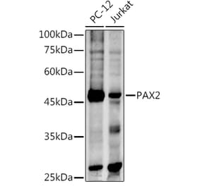
![Western Blot - Anti-PAX2 Antibody [PAX2/1105] (A277731) - Antibodies.com](https://cdn.antibodies.com/image/catalog/277/A277731_1.jpg?profile=product_alternative)
![Western Blot - Anti-PAX2 Antibody [PAX2/1105] - BSA and Azide free (A278319) - Antibodies.com](https://cdn.antibodies.com/image/catalog/278/A278319_1.jpg?profile=product_alternative)