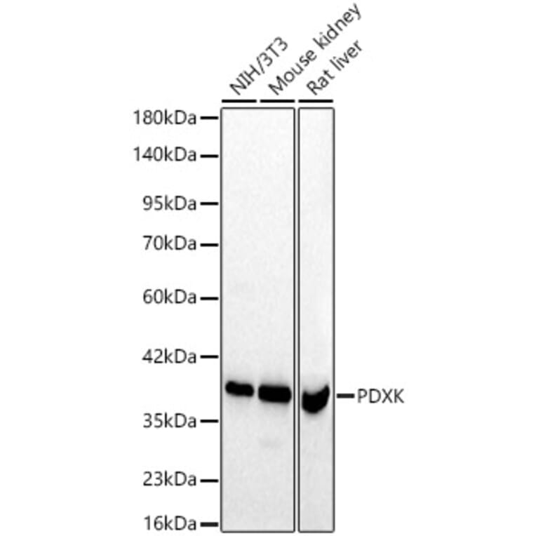
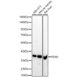
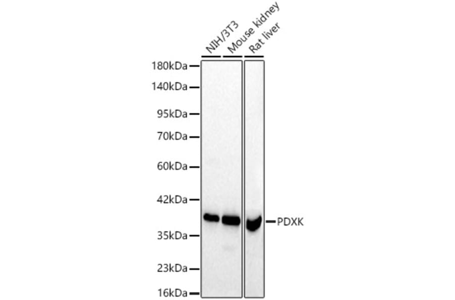
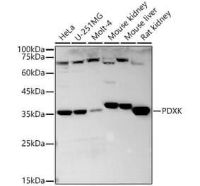
PDXK.1 expression in various lysates analyzed by western blot. Primary antibody incubation was performed for 1 hour on 25ug protein per lane with Anti-PDXK.1 Antibody (A329731) at a dilution of 1:1000 and detected with chemiluminescence.
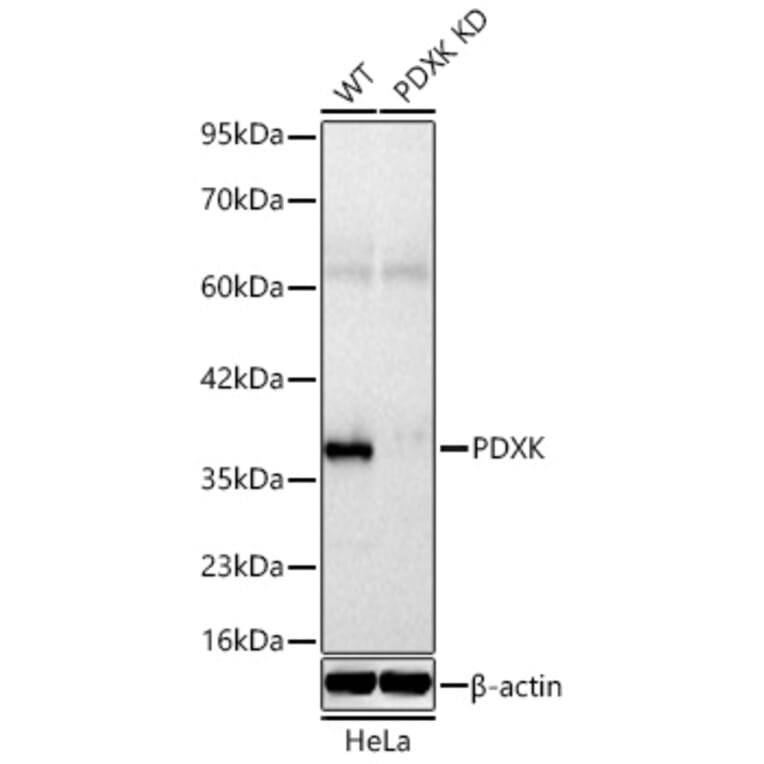
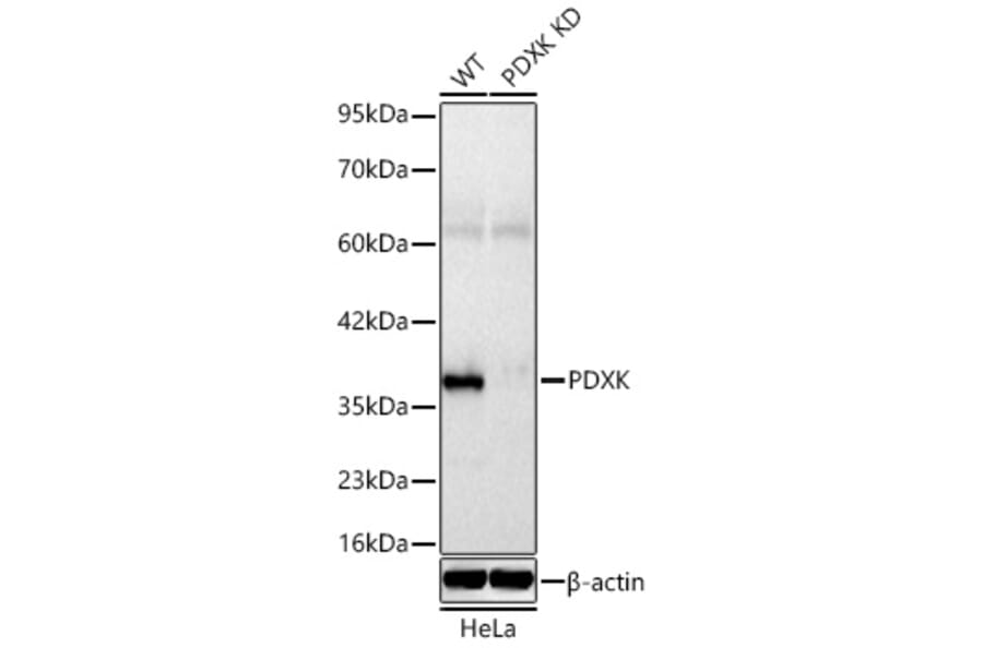
PDXK.1 expression in lysates from wild type (WT) and PDXK knockdown (KD) HeLa cells analyzed by western blot. Primary antibody incubation was performed for 1 hour on 25ug protein per lane with Anti-PDXK.1 Antibody (A329731) at a dilution of 1:1600 and detected with chemiluminescence.
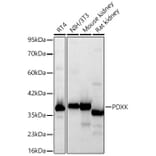
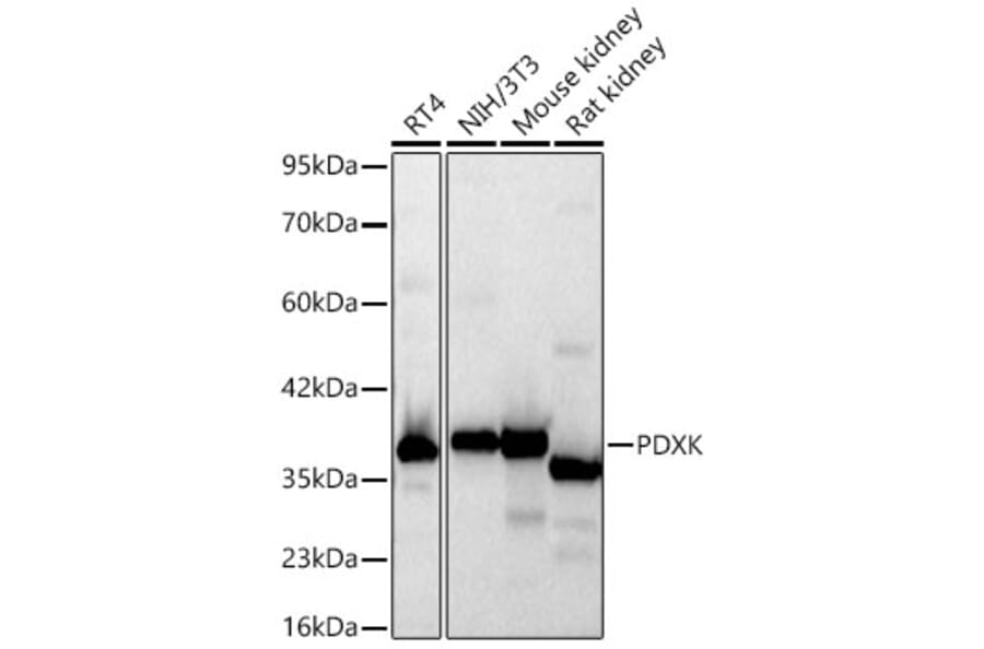
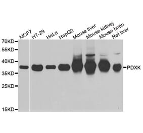
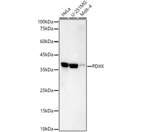
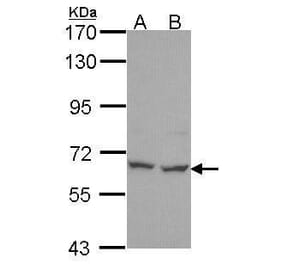
PDXK.1 expression in various lysates analyzed by western blot. Primary antibody incubation was performed for 1 hour on 25ug protein per lane with Anti-PDXK.1 Antibody (A329731) at a dilution of 1:1600 and detected with chemiluminescence.
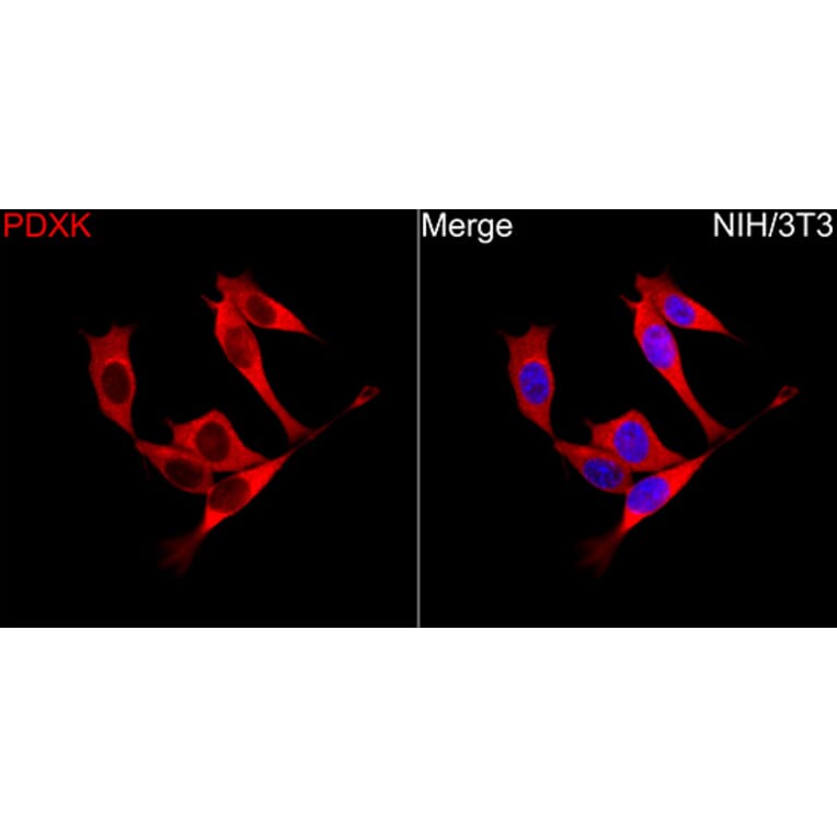
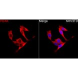
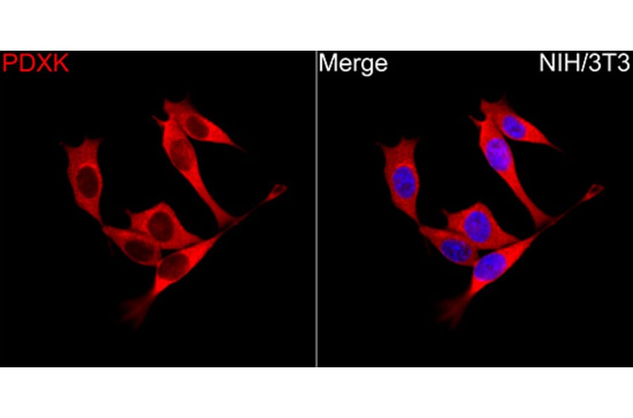
PDXK.1 expression in NIH/3T3 cells analyzed by immunofluorescence. Staining was performed with Anti-PDXK.1 Antibody (A329731) at a dilution of 1:100 followed by Cy3-conjugated Goat anti-Rabbit IgG (H+L) secondary antibody at a dilution of 1:500. Nuclei were stained with DAPI (blue).
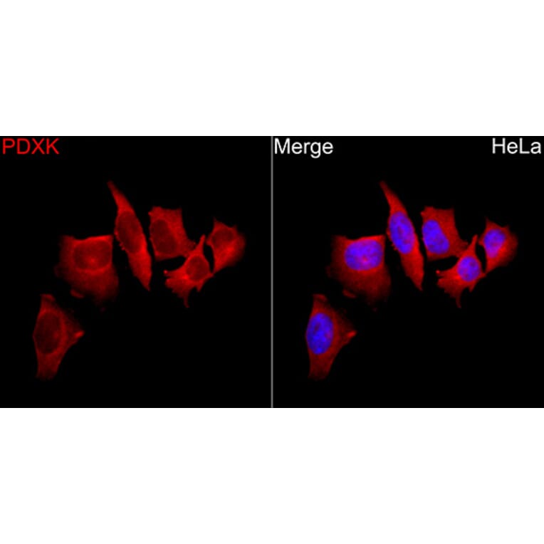
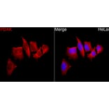
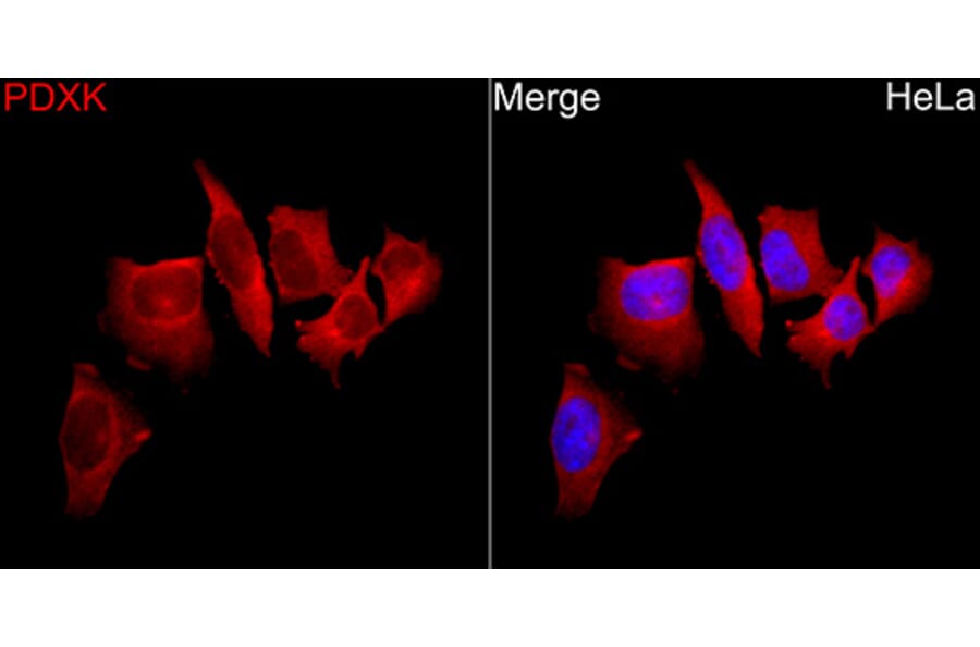
PDXK.1 expression in Hela cells analyzed by immunofluorescence. Staining was performed with Anti-PDXK.1 Antibody (A329731) at a dilution of 1:100 followed by Cy3-conjugated Goat anti-Rabbit IgG (H+L) secondary antibody at a dilution of 1:500. Nuclei were stained with DAPI (blue).