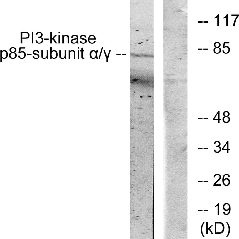
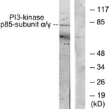
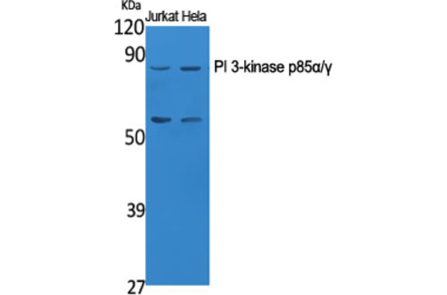
Western Blot - Anti-PI 3 Kinase p85 alpha + Gamma Antibody (A93926)
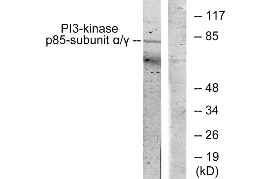
Western blot analysis of lysates from COS7 cells, treated with H2O2 100uM 30' using Anti-PI 3 Kinase p85 alpha + Gamma Antibody. The right hand lane represents a negative control, where the antibody is blocked by the immunising peptide.
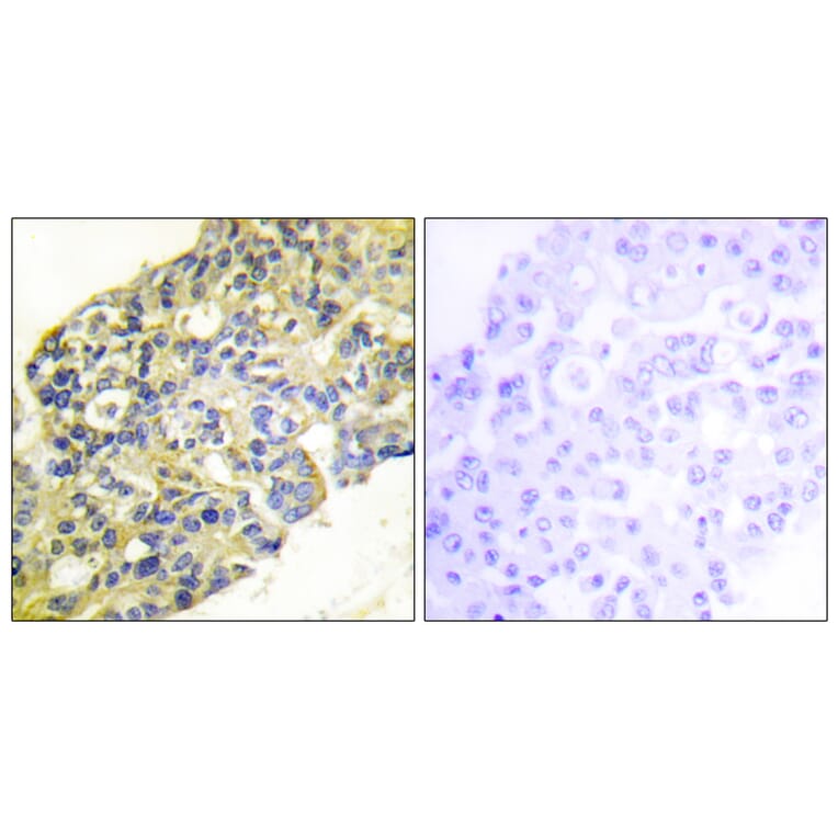
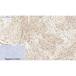
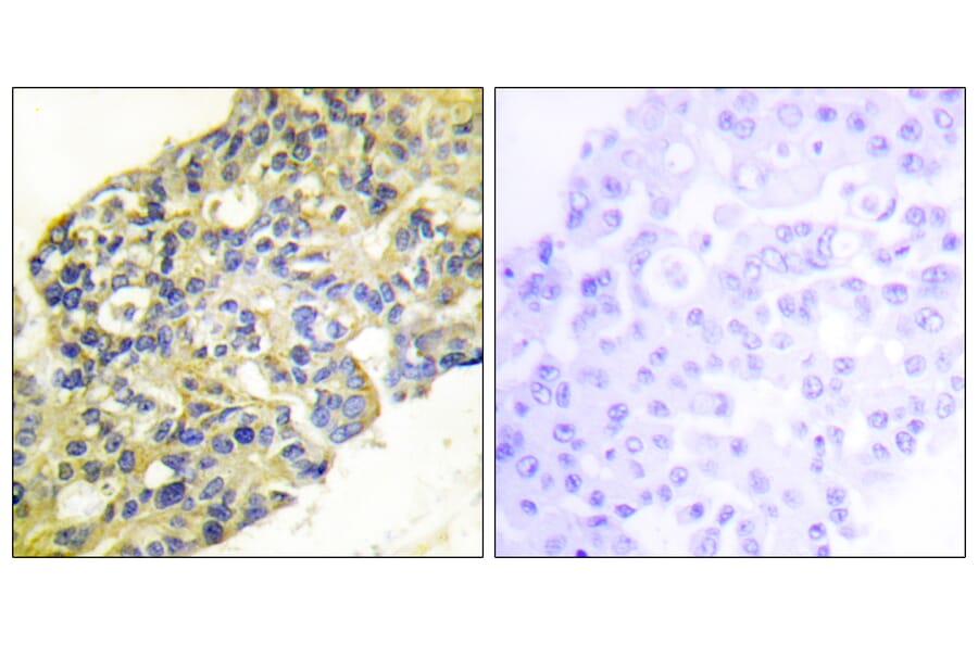
Immunohistochemical analysis of paraffin-embedded human breast carcinoma tissue using Anti-PI 3 Kinase p85 alpha + Gamma Antibody. The right hand panel represents a negative control, where the antibody was pre-incubated with the immunising peptide.
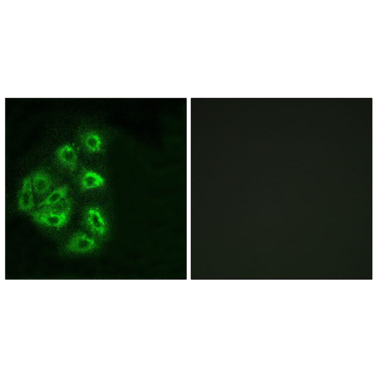
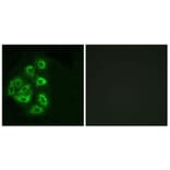
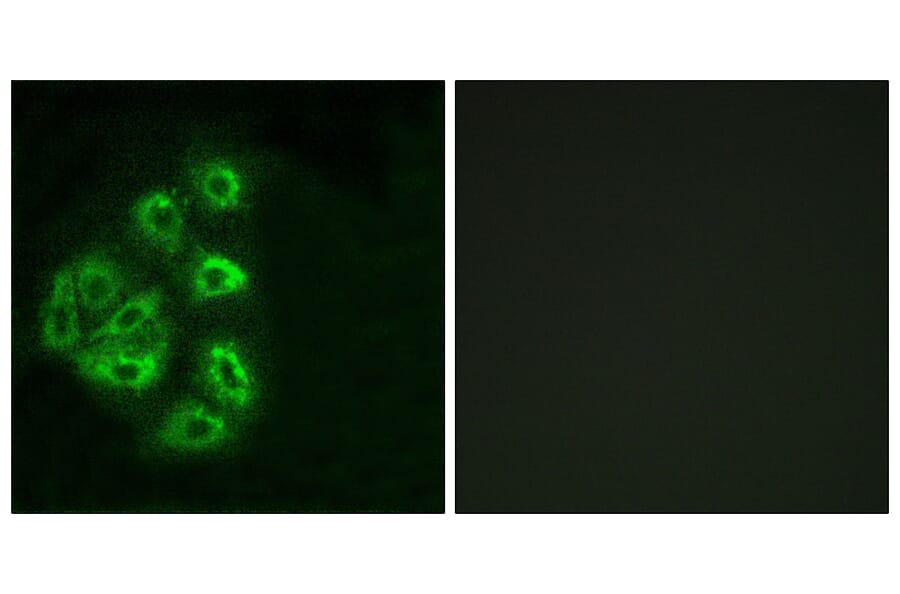
Immunofluorescence analysis of HeLa cells using Anti-PI 3 Kinase p85 alpha + Gamma Antibody. The right hand panel represents a negative control, where the antibody was pre-incubated with the immunising peptide.
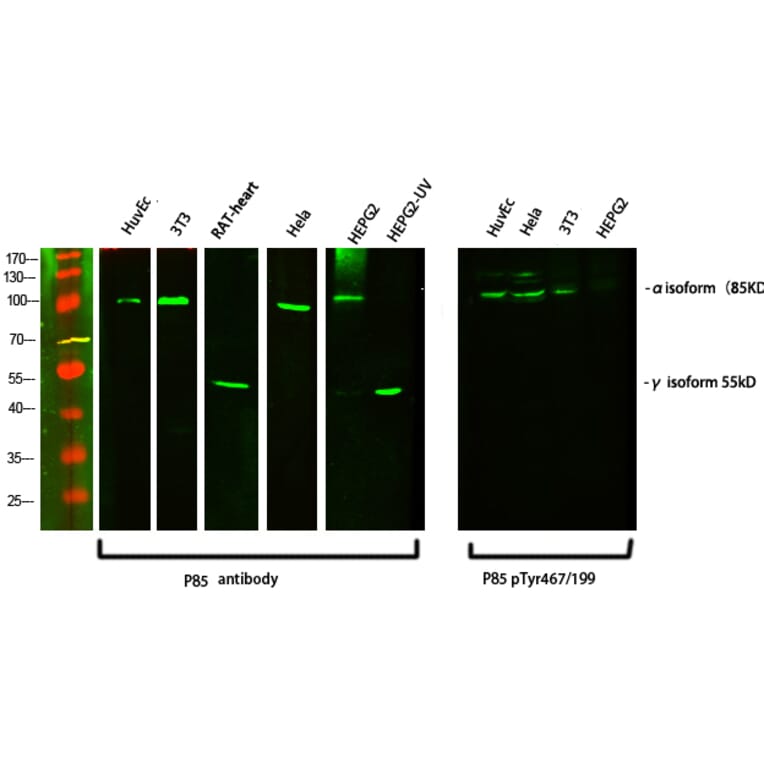
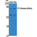
Western Blot - Anti-PI 3 Kinase p85 alpha + Gamma Antibody (A93926)
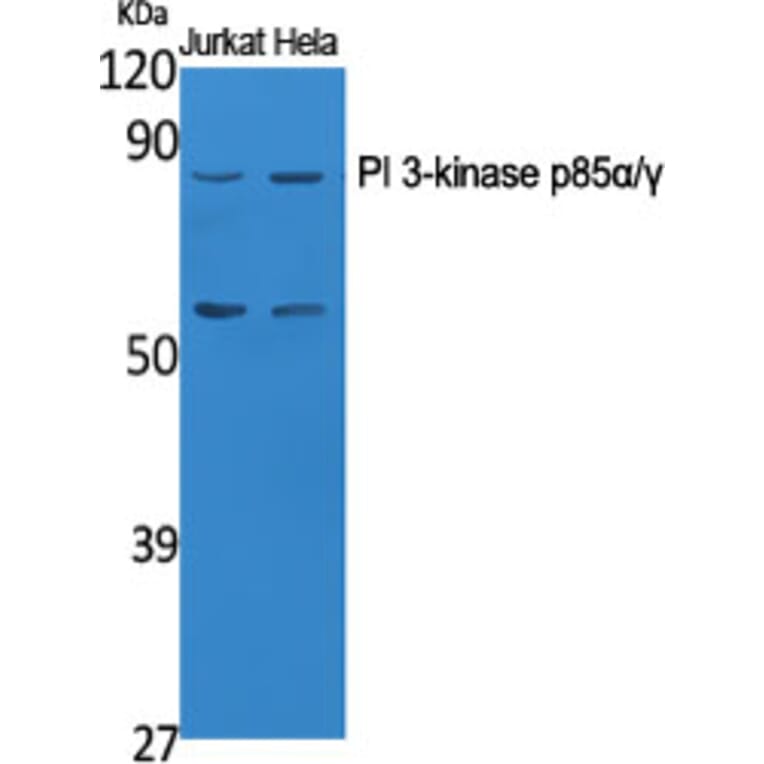
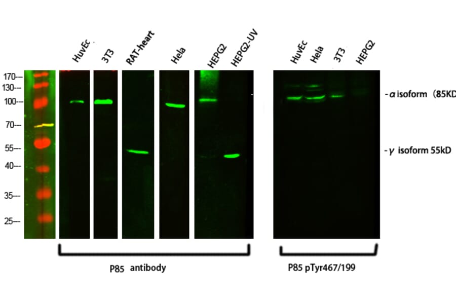
Western blot analysis of various cells using Anti-PI 3 Kinase p85 alpha + Gamma Antibody at 1:1,000 (4°C overnight). Goat Anti-Rabbit IgG (IRDye 800) was used as a secondary at 1:5,000 (25°C, 1 hour).
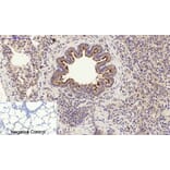
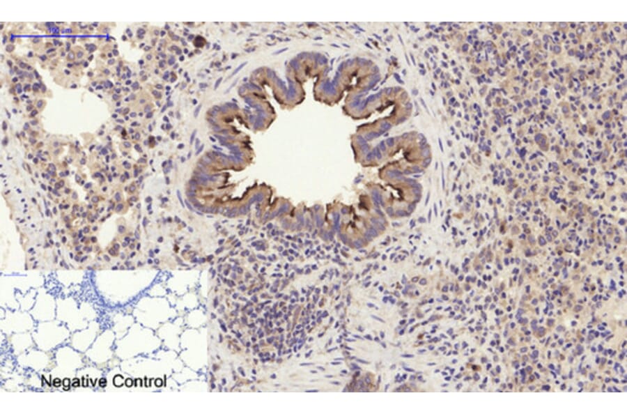
Immunohistochemical analysis of paraffin-embedded human uterus tissue using Anti-PI 3 Kinase p85 alpha + Gamma Antibody at 1:200 (4°C overnight). Negative control was secondary antibody only.
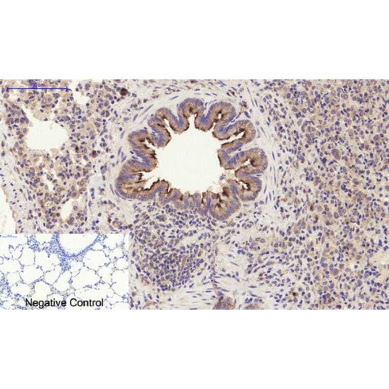
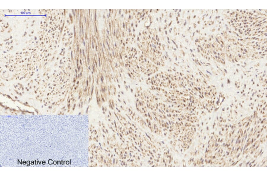
Immunohistochemical analysis of paraffin-embedded rat lung tissue using Anti-PI 3 Kinase p85 alpha + Gamma Antibody at 1:200 (4°C overnight). Negative control was secondary antibody only.
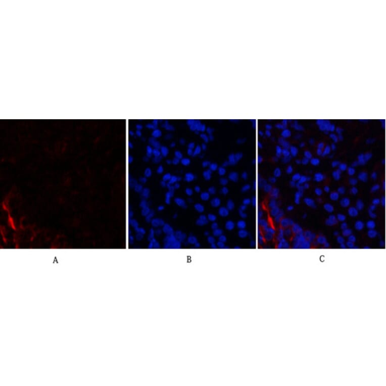
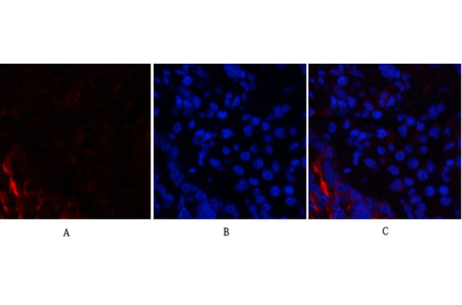
Immunofluorescence analysis of rat lung tissue using Anti-PI 3 Kinase p85 alpha + Gamma Antibody (red) at 1:200 (4°C overnight). Cy3 labelled secondary antibody was used at 1:300 (RT 50min). Panel A: Target. Panel B: DAPI. Panel C: Merge.
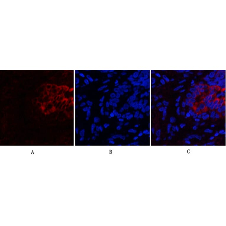
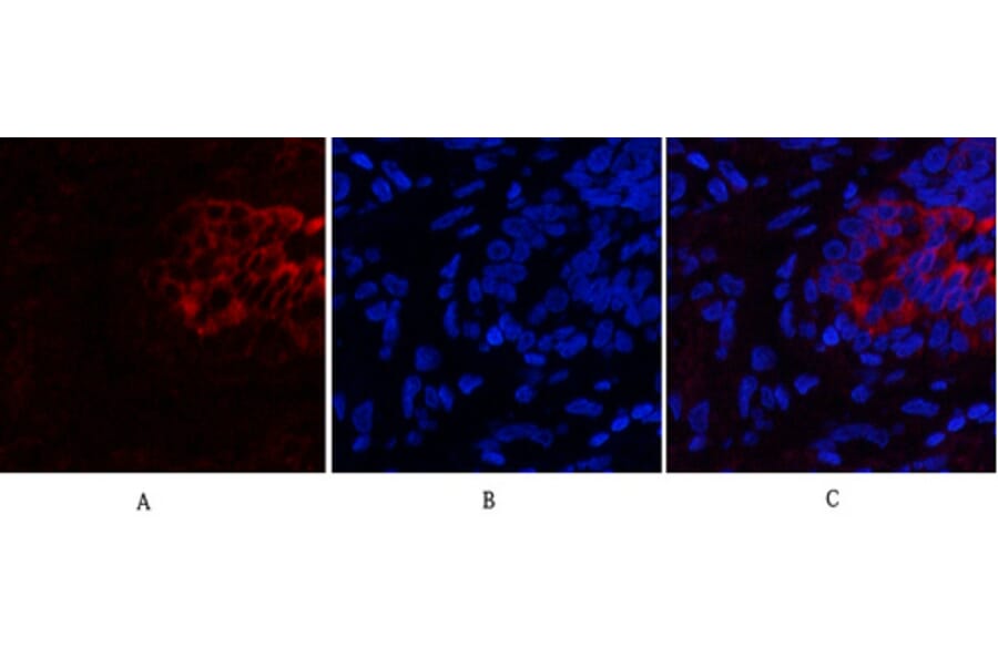
Immunofluorescence analysis of human lung tissue using Anti-PI 3 Kinase p85 alpha + Gamma Antibody (red) at 1:200 (4°C overnight). Cy3 labelled secondary antibody was used at 1:300 (RT 50min). Panel A: Target. Panel B: DAPI. Panel C: Merge.