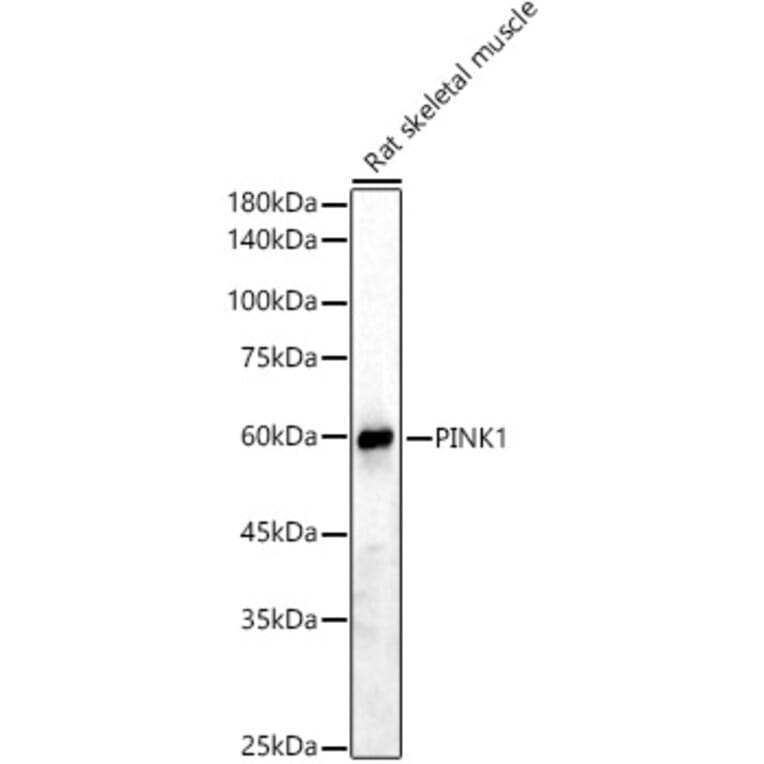
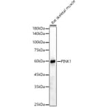
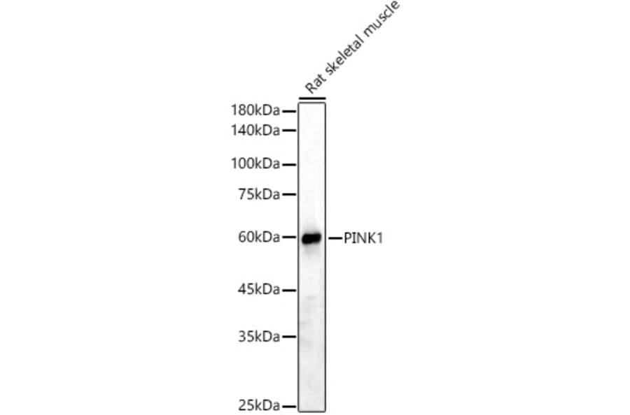
Western blot analysis of Rat skeletal muscle, using Anti-PINK1 Antibody (A15557) at 1:2,000 dilution. The secondary antibody was Goat Anti-Rabbit IgG H&L Antibody (HRP) at 1:10,000 dilution. Lysates/proteins were present at 25µg per lane. The blocking buffer used was 3% non-fat dry milk in TBST. Detection was with a ECL Basic Kit. Exposure time: 90s.
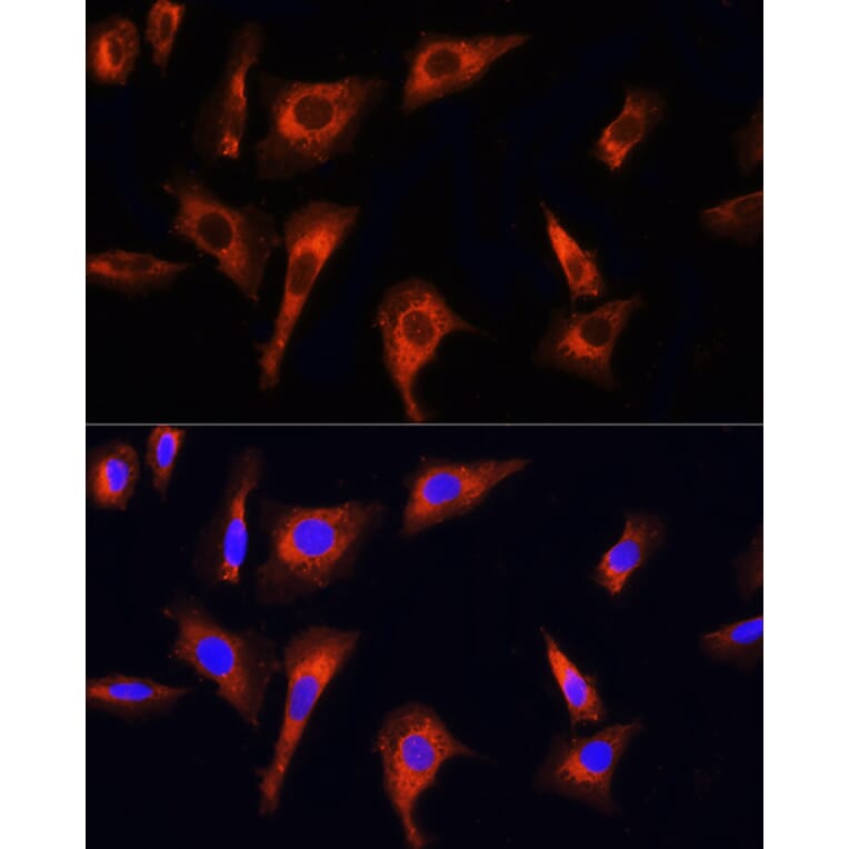
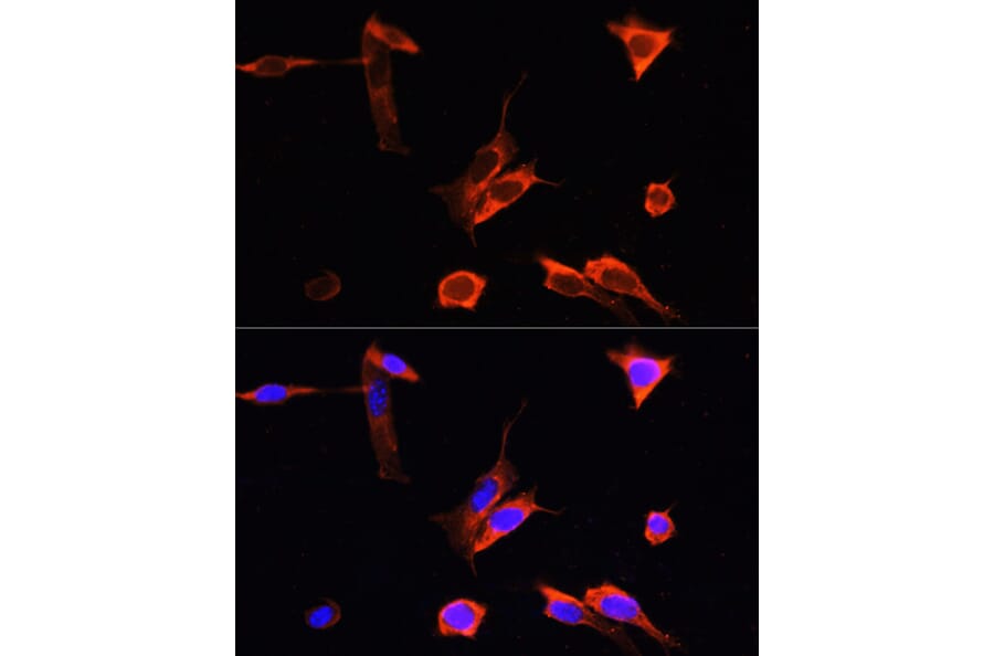
Immunofluorescence analysis of NIH-3T3 cells using Anti-PINK1 Antibody (A15557) at dilution of 100 (40x lens). DAPI was used to stain the cell nuclei (blue).
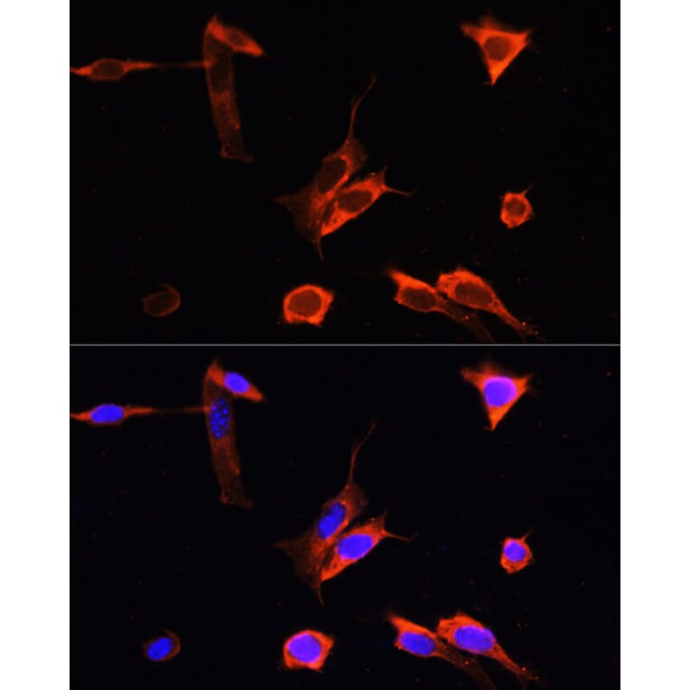
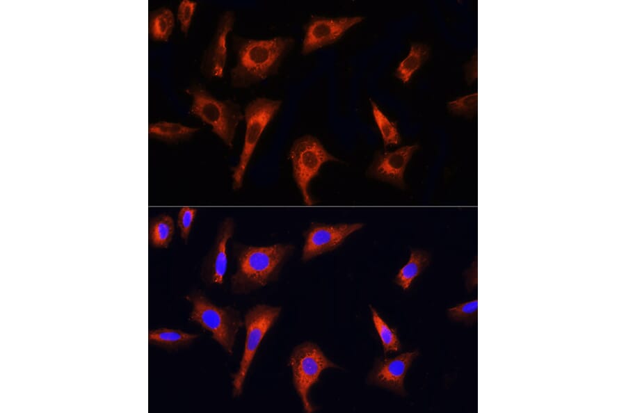
Immunofluorescence analysis of U-2 OS cells using Anti-PINK1 Antibody (A15557) at dilution of 100 (40x lens). DAPI was used to stain the cell nuclei (blue).
Publishing research using Anti-PINK1 Antibody (A15557)? Please let us know so that we can list the citation on this page.
Alternative products to Anti-PINK1 Antibody (A15557)


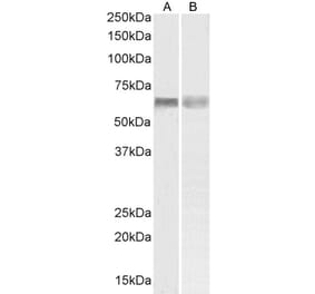
![Western Blot - Anti-PINK1 Antibody [S4-15] (A305003) - Antibodies.com](https://cdn.antibodies.com/image/catalog/305/A305003_1.png?profile=product_alternative)