Supplied in Phosphate Buffered Saline, pH 7.3, with 50% Glycerol, 0.05% BSA, and 0.02% Sodium Azide.
Storage
Shipped at 4°C. Upon delivery aliquot and store at -20°C. Avoid freeze/thaw cycles.
Synonyms
CAL, CFTR-associated ligand, FIG, Fused in glioblastoma, Golgi-associated PDZ and coiled-coil motif-containing protein, GOPC, PDZ protein interacting specifically with TC10
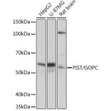
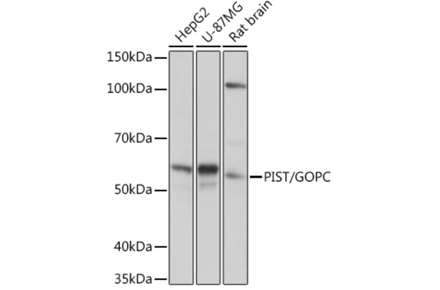
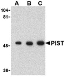
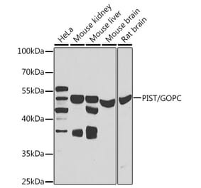
Western Blot - Anti-PIST Antibody [ARC1831] (A307711)
Western blot analysis of extracts of various cell lines, using Anti-PIST Antibody (A307711) at 1:1,000 dilution. The secondary antibody was Goat Anti-Rabbit IgG H&L Antibody (HRP) at 1:10,000 dilution. Lysates/proteins were present at 25µg per lane. The blocking buffer used was 3% non-fat dry milk in TBST. Detection was with a ECL Basic Kit. Exposure time: 1s.
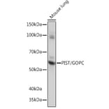
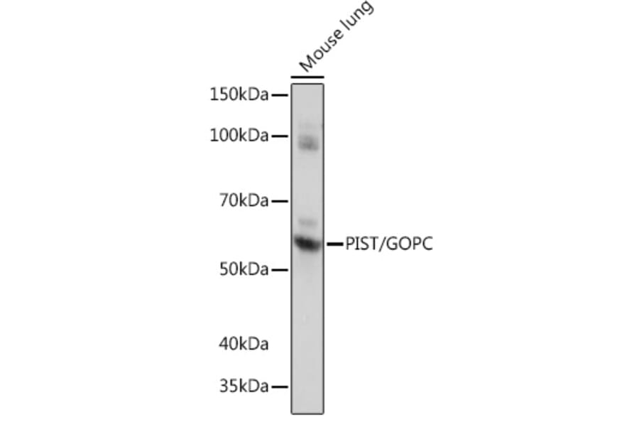
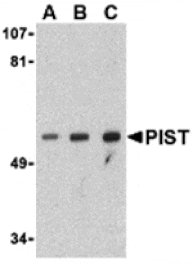
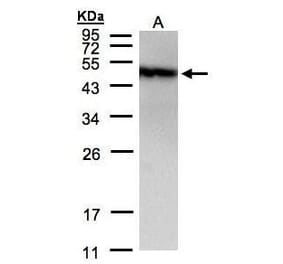
Western Blot - Anti-PIST Antibody [ARC1831] (A307711)
Western blot analysis of extracts of Mouse lung, using Anti-PIST Antibody (A307711) at 1:1,000 dilution. The secondary antibody was Goat Anti-Rabbit IgG H&L Antibody (HRP) at 1:10,000 dilution. Lysates/proteins were present at 25µg per lane. The blocking buffer used was 3% non-fat dry milk in TBST. Detection was with a ECL Basic Kit. Exposure time: 5s.
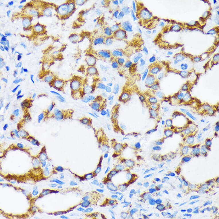
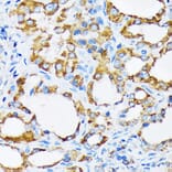
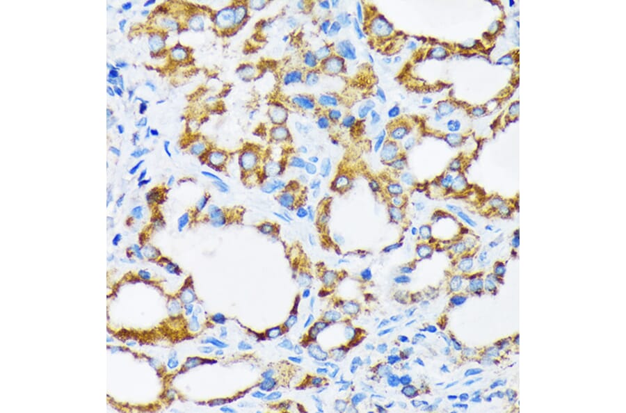
Immunohistochemistry analysis of paraffin-embedded human thyroid cancer using Anti-PIST Antibody (A307711) at a dilution of 1:100 (40x lens). Perform microwave antigen retrieval with 10 mM Tris/EDTA buffer pH 9.0 before commencing with IHC staining protocol.
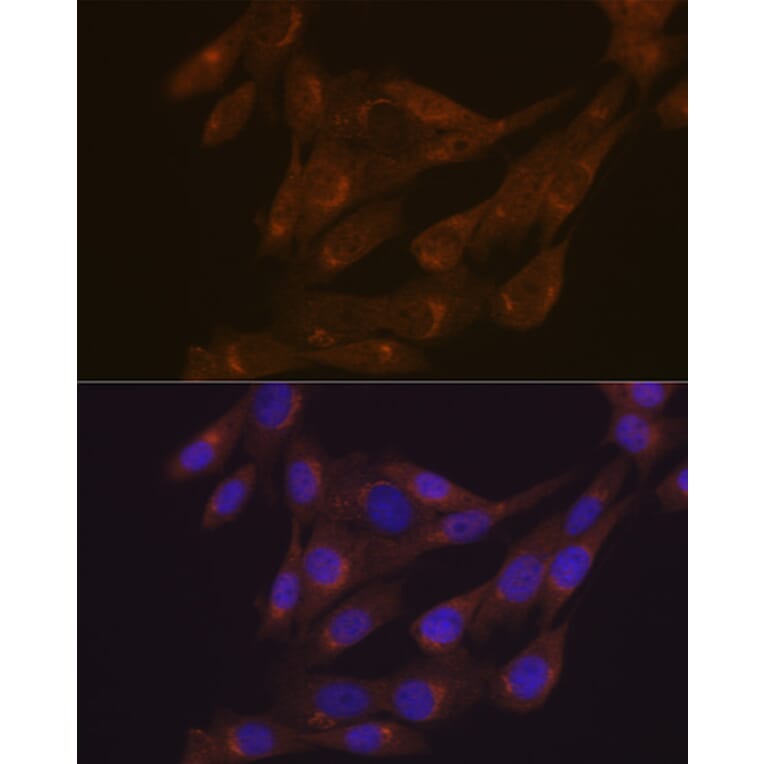
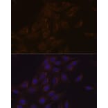
Immunofluorescence analysis of C6 cells using Anti-PIST Antibody (A307711) at a dilution of 1:100 (40x lens). DAPI was used to stain the cell nuclei (blue).
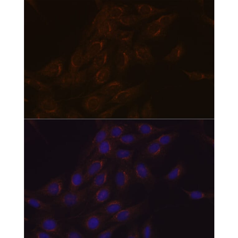
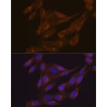
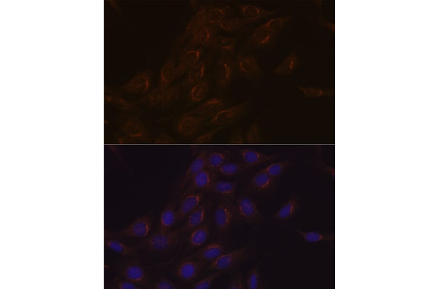
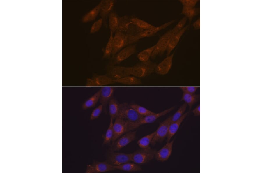
Immunofluorescence analysis of NIH-3T3 cells using Anti-PIST Antibody (A307711) at a dilution of 1:100 (40x lens). DAPI was used to stain the cell nuclei (blue).