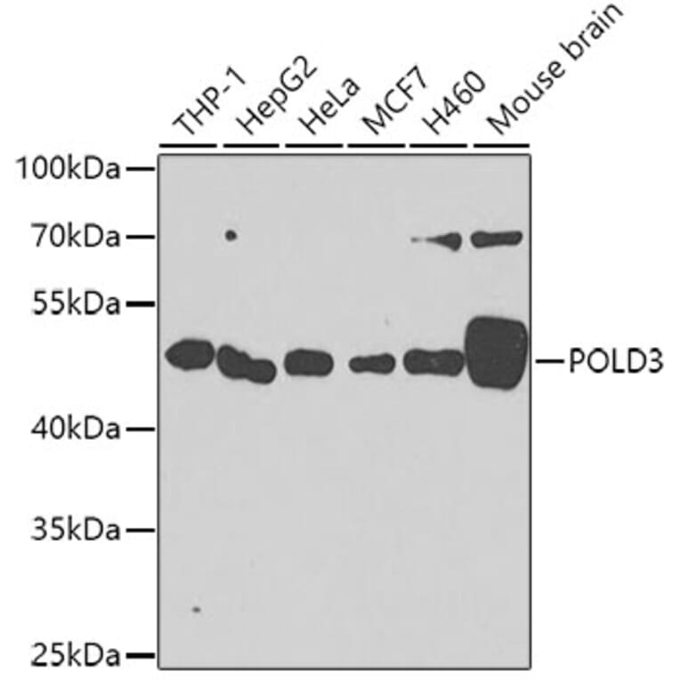
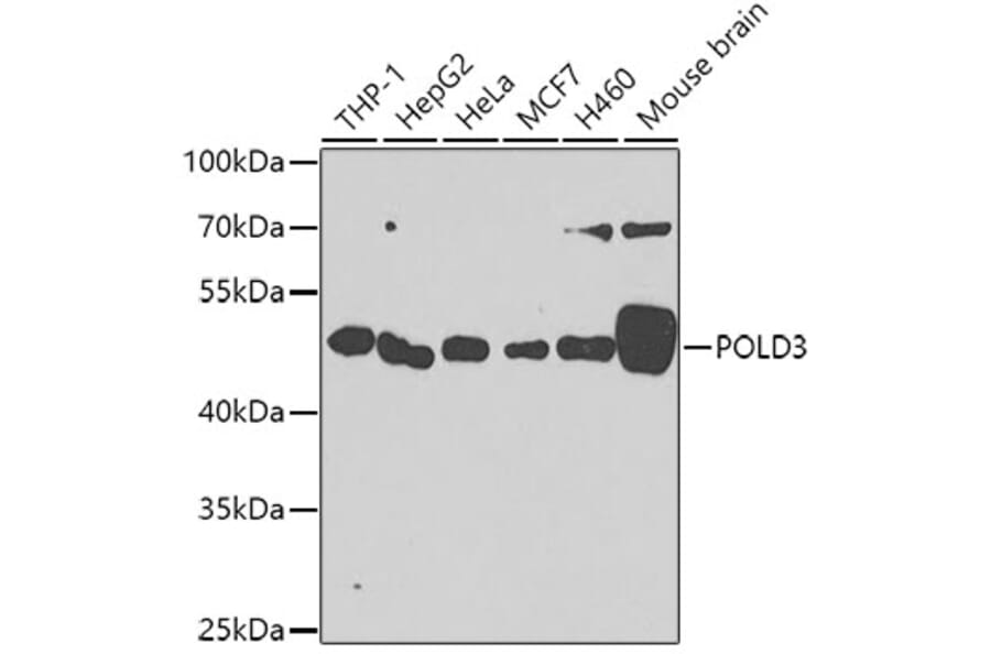
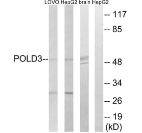
Western blot analysis of extracts of various cell lines, using Anti-POLD3 Antibody (A10090) at 1:1,000 dilution. The secondary antibody was Goat Anti-Rabbit IgG H&L Antibody (HRP) at 1:10,000 dilution. Lysates/proteins were present at 25µg per lane. The blocking buffer used was 3% non-fat dry milk in TBST. Detection was with a ECL Enhanced Kit. Exposure time: 60s.
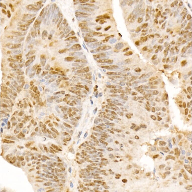
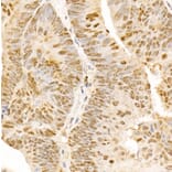
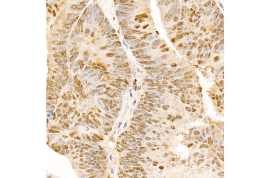
Immunohistochemistry analysis of paraffin-embedded human colon carcinoma tissue using Anti-POLD3 Antibody (A10090) at a dilution of 1:300 (40x lens). Perform microwave antigen retrieval with 10 mM PBS buffer pH 7.2 before commencing with IHC staining protocol.
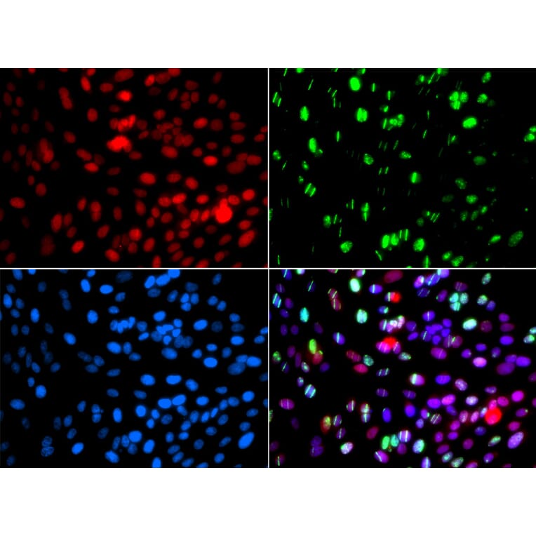
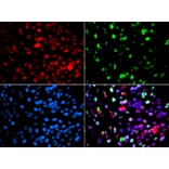
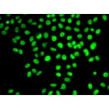
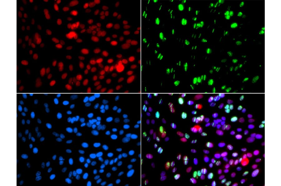
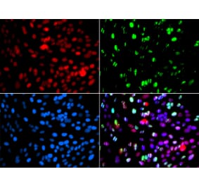
Immunofluorescence analysis of GFP-RNF168 transgenic U2OS cells using Anti-POLD3 Antibody (A10090). Green:GFP-RNF168 fusion protein expression for DNA damage marker. DAPI was used to stain the cell nuclei (blue). RNF168(GFP) can be used to mark cells damaged by UV-A laser for they always gather around DNA damage region.