This antibody targets the C-terminal region of human PTH1R, recognizing the non-phosphorylated form. It is suitable for Western blot analysis, enables immunoprecipitation from cell and tissue lysates, and is optimized for immunohistochemistry in cultured cells and tissue sections.
Applications
WB, ICC, IHC
Dilutions
WB: 1,000, IHC: 1:100, ICC: 1:200
Reactivity
Human
Immunogen
Synthetic peptide corresponding to the C-terminal of human PTH1R (amino acids. 473-493). Immunogen range is 22-22 amino acids.
Sequence
EEASGPERPPALLQEEWETVM
Host
Rabbit
Clonality
Polyclonal
Isotype
IgG
Conjugate
Unconjugated
Purification
Antigen affinity purification.
Concentration
Lot Specific
Product Form
Liquid
Formulation
Supplied in Dulbecco's PBS, pH 7.4, with 150 mM NaCl and 0.005% Sodium Azide.
Storage
Shipped at 4°C. Upon delivery aliquot and store at -20°C. Avoid freeze/thaw cycles.
Synonyms
Parathyroid hormone 1 receptor, Parathyroid hormone/parathyroid hormone-related peptide receptor, PTH/PTHr receptor, PTH/PTHrP type I receptor, PTH1 receptor, PTHR, PTHR1
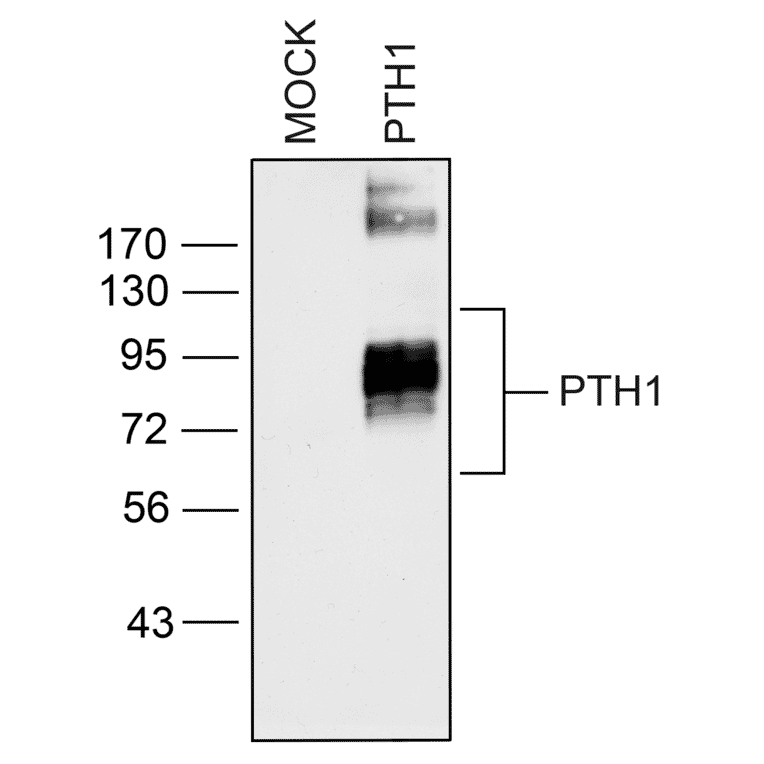
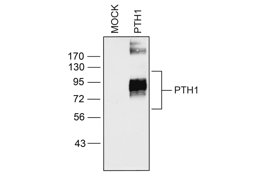
Western blot validation of Parathyroid Hormone Receptor 1 (PTH1R) in transfected HEK293 cells. Lysates from native HEK293 cells (MOCK) or cells stably expressing PTH1R were immunoblotted with anti-PTH1R antibody (A334534) at 1:1000.
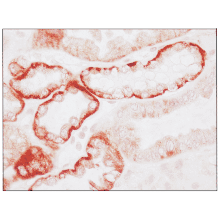
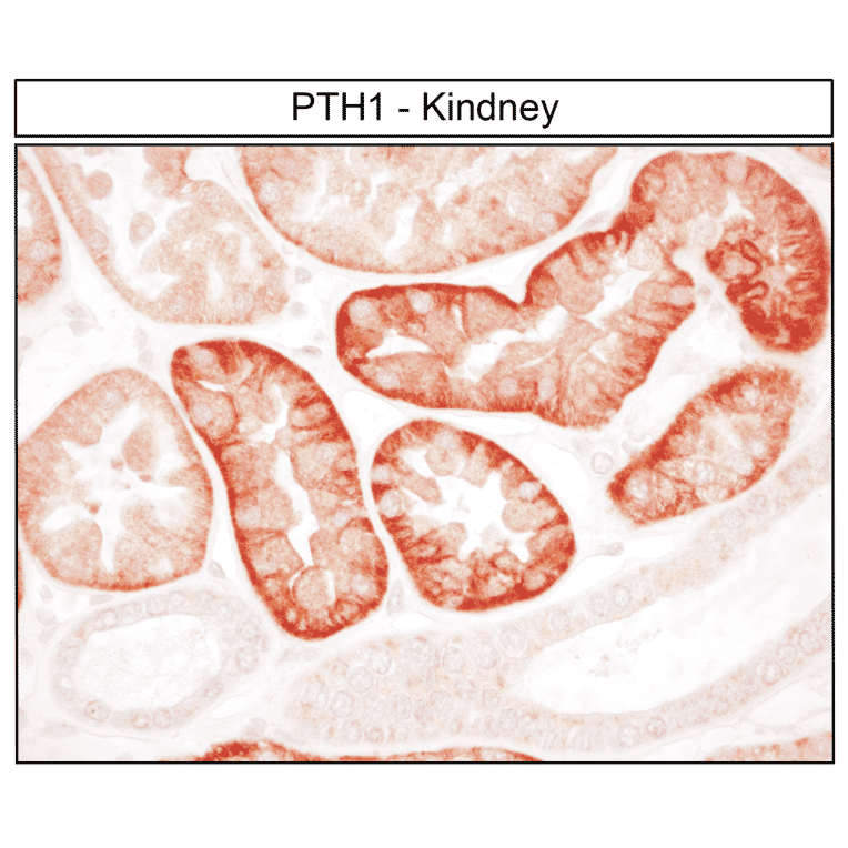
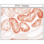
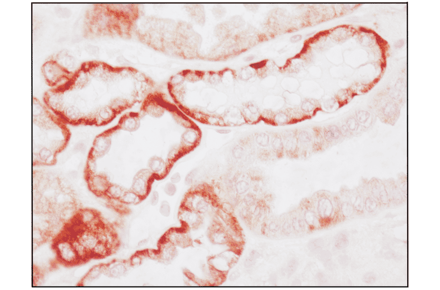
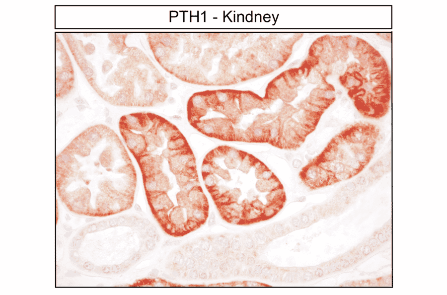
Immunohistochemical detection of Parathyroid Hormone Receptor 1 (PTH1R) in human kidney. Kidney sections were dewaxed, microwaved in citric acid, and stained with anti-PTH1R antibody (A334534) at 1:100, followed by biotinylated anti-rabbit IgG and avidin-biotin solution. Sections were developed with 3,3-diaminobenzidine (DAB)-glucose oxidase and lightly counterstained with hematoxylin. PTH1R receptors were uniformly detected at the basolateral plasma membrane of tubular cells.
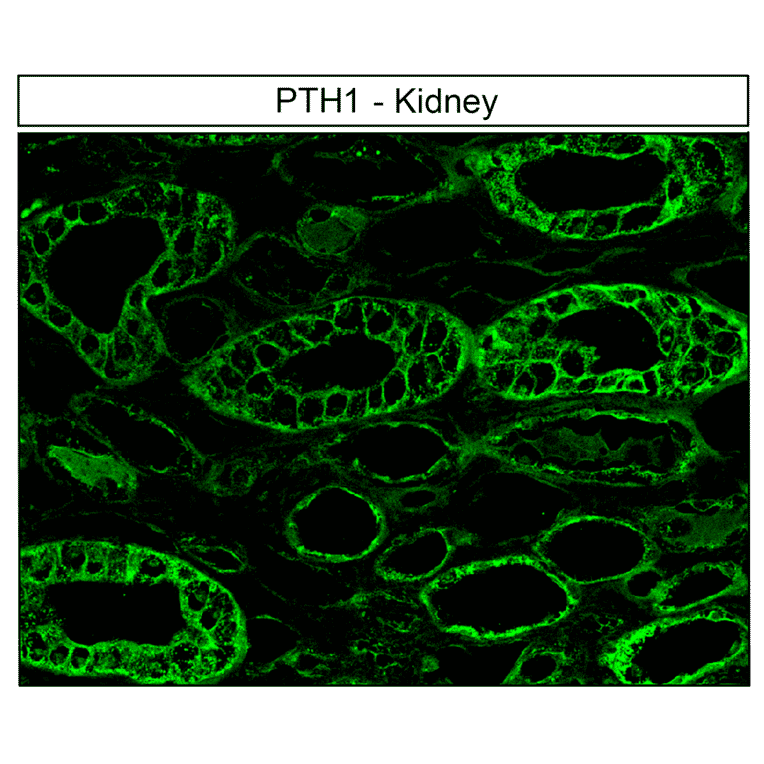
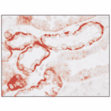
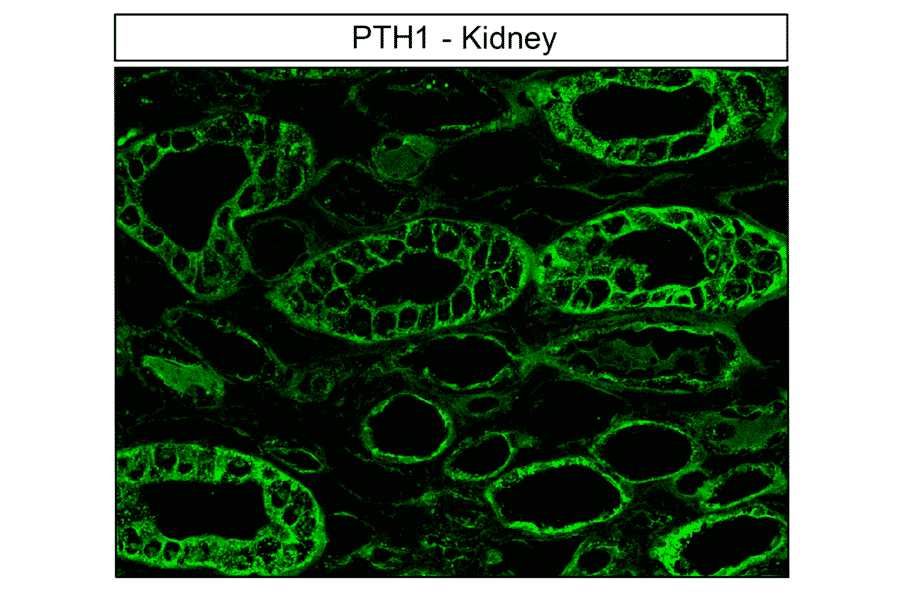
Immunofluorescent localization of Parathyroid Hormone Receptor 1 (PTH1R) in human kidney distal convoluted tubules and connecting ducts. Kidney sections were dewaxed, microwaved in citric acid, and incubated with anti-PTH1R antibody (A334534) at 1:100, followed by anti-rabbit-AlexaFluor488. PTH1R receptors were detected at the plasma membrane of all cells.
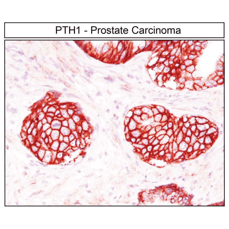
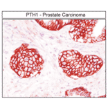
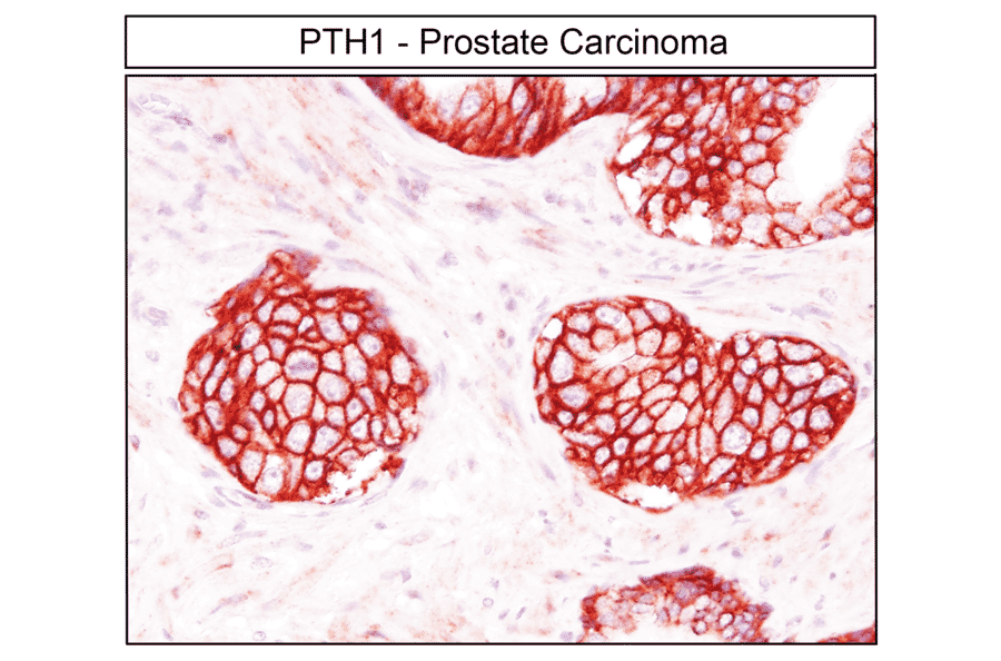
Immunohistochemical localization of Parathyroid Hormone Receptor 1 (PTH1R) in human prostate carcinoma. Prostate carcinoma sections were dewaxed, microwaved in citric acid, and stained with anti-PTH1R antibody (A334534) at 1:100, followed by biotinylated anti-rabbit IgG and avidin-biotin solution. Color was developed with 3-amino-9-ethylcarbazole (AEC), and sections were counterstained with hematoxylin. PTH1R receptors were detected at the plasma membrane of nearly all tumor cells.
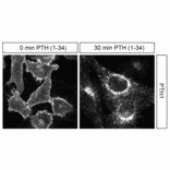
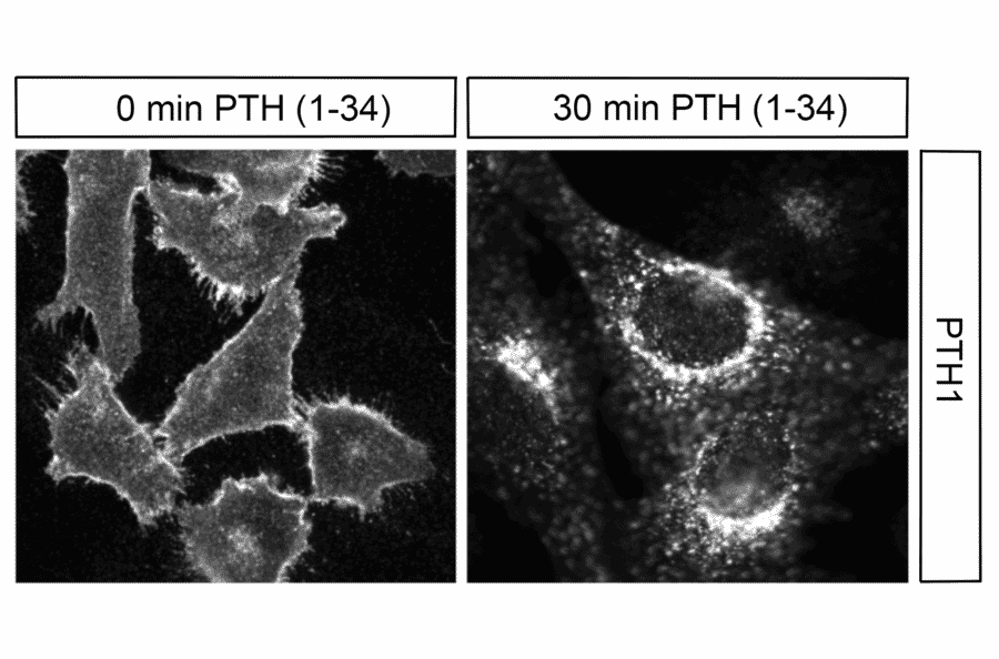
Immunocytochemical analysis of Parathyroid Hormone Receptor 1 (PTH1R) in HEK293 cells. HEK293 cells stably expressing PTH1R were untreated or treated with 1 µM PTH 1-34 for 30 min and stained with anti-PTH1R antibody (A334534) at 1:200. PTH1R receptors were localized to the plasma membrane in untreated cells (0 min) and in perinuclear vesicular clusters after 30 min of PTH exposure.
Detection of Parathyroid Hormone Receptor 1 (PTH1R) in human kidney by immunohistochemistry. Kidney sections were dewaxed, microwaved in citric acid, and incubated with anti-PTH1R antibody (A334534) at 1:100, followed by biotinylated anti-rabbit IgG and avidin-biotin solution. Sections were developed with 3,3-diaminobenzidine (DAB)-glucose oxidase and lightly counterstained with hematoxylin. PTH1R receptors were uniformly observed at the basolateral plasma membrane of tubular cells.
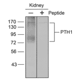
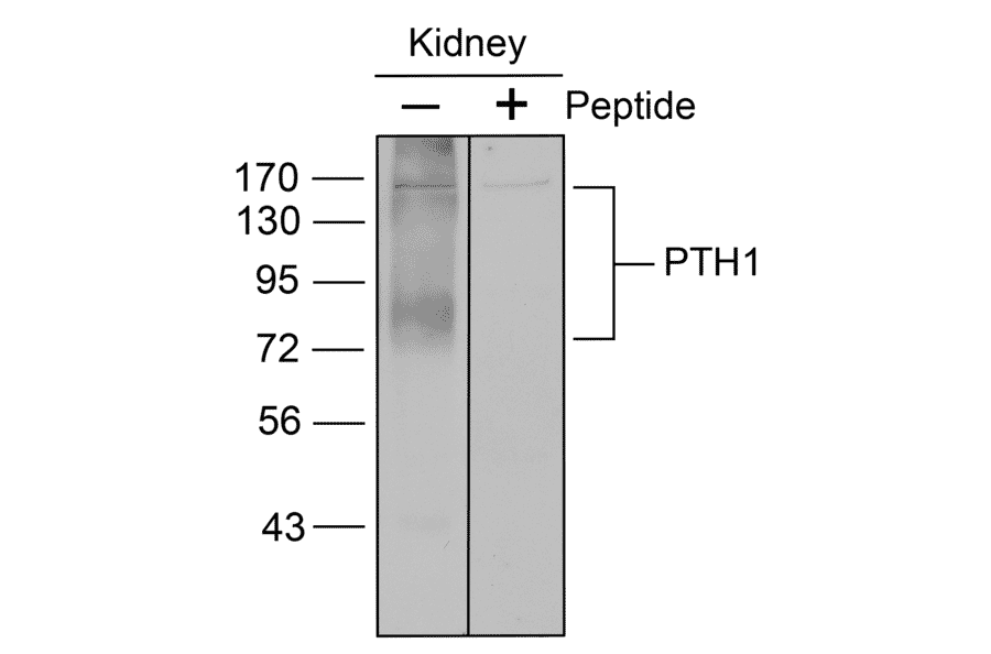
Western blot analysis of Parathyroid Hormone Receptor 1 (PTH1R) in human kidney membrane preparations. Kidney membrane samples were separated on 8% SDS-polyacrylamide gels, blotted onto nitrocellulose, and probed with anti-PTH1R antibody (A334534) at 1:1000 in the presence (+) or absence (-) of 10 µg/ml antigen peptide. PTH1R receptors were detected only in the absence of the antigen peptide.


![SDS-PAGE - Anti-PTH1R Nanobody [SAA0797] (A337688) - Antibodies.com](https://cdn.antibodies.com/image/catalog/337/A337688_1.jpg?profile=product_alternative)
![SDS-PAGE - Anti-PTH1R Nanobody [SAA1275] (A337936) - Antibodies.com](https://cdn.antibodies.com/image/catalog/337/A337936_1.jpg?profile=product_alternative)