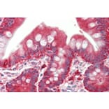
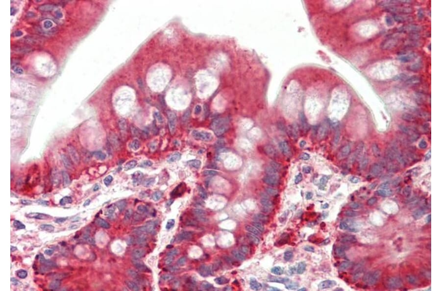
PYROXD1 expression in Human Small Intestine analyzed by immunohistochemistry. Tissue was paraffin-embedded, and antigen retrieval was achieved by steaming in citrate buffer, pH 6. Staining was performed with Anti-PYROXD1 Antibody (A85088) at 5µg/ml and revealed with alkaline phosphatase (AP).
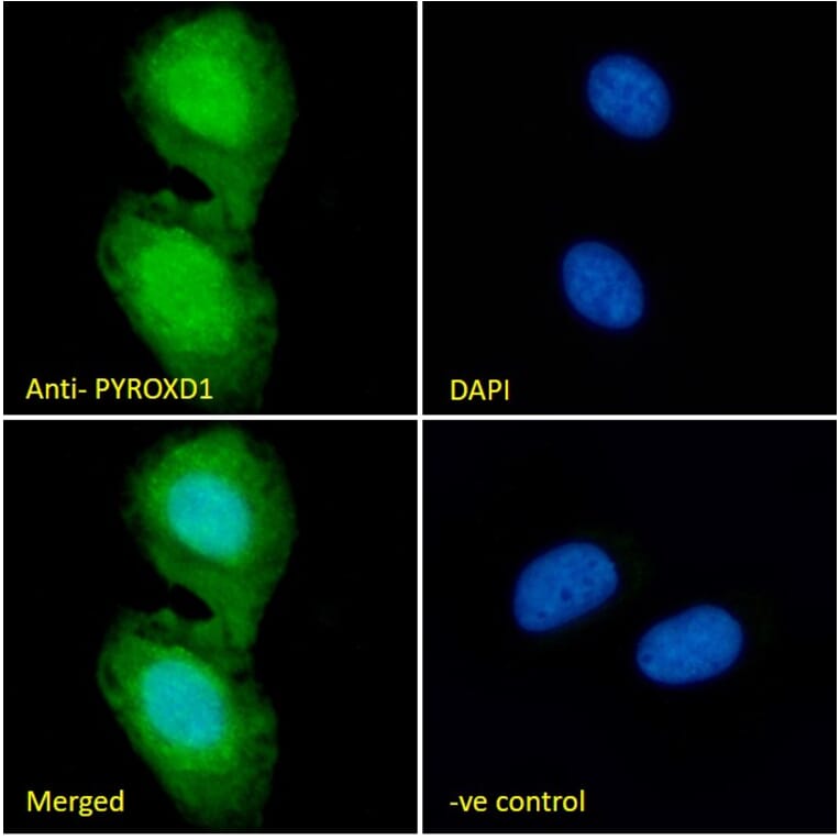
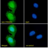
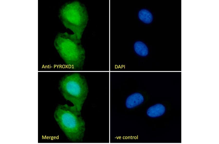
PYROXD1 expression in U2OS cells analyzed by immunofluorescence. Cells were permeabilized with 0.15% Triton. Staining was performed with Anti-PYROXD1 Antibody (A85088) at 10µg/ml for 1 hour and Alexa Fluor 488 secondary antibody at 2µg/ml. Nuclear and weak cytoplasmic staining shown and nuclei were stained with DAPI (blue). Negative control: Goat IgG Isotype Control at 10µg/ml followed by Alexa Fluor 488 secondary antibody at 2µg/ml.
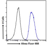
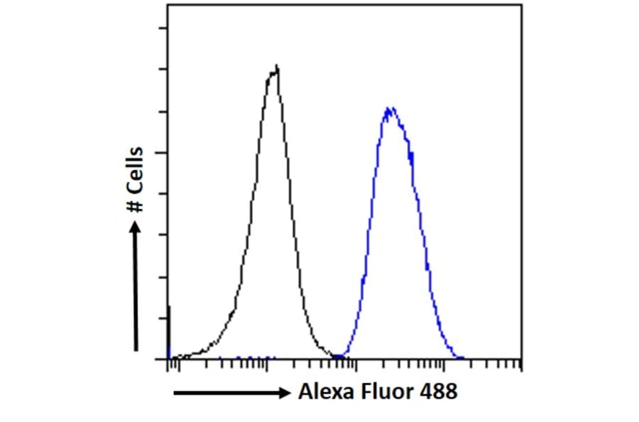
PYROXD1 expression in U2OS cells (blue line) analyzed by flow cytometry. Cells were fixed in PFA and permeabilized with 0.5% Triton. Staining was performed with Anti-PYROXD1 Antibody (A85088) at 10µg/ml for 1 hour and Alexa Fluor 488 secondary antibody at 1µg/ml. Negative Control: Goat IgG Isotype Control (black line) followed by Alexa Fluor 488 secondary antibody.
Publishing research using Anti-PYROXD1 Antibody (A85088)? Please let us know so that we can list the citation on this page.
Proteins predicted to interact with PYROXD1
Predicted protein interactions based upon String database. Revelancy score correlates with probability of interaction.