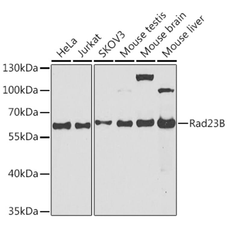
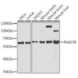
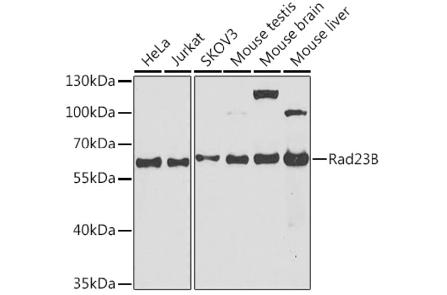
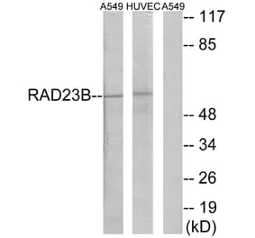
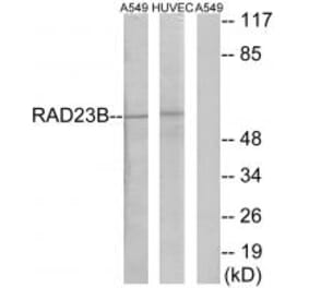
RAD23B expression in various lysates analyzed by western blot. Primary antibody incubation was performed for 1 hour on 25ug protein per lane with Anti-RAD23B Antibody (A12901) at a dilution of 1:1000 and detected with chemiluminescence.
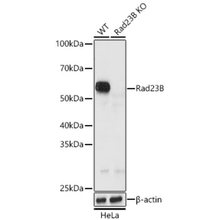
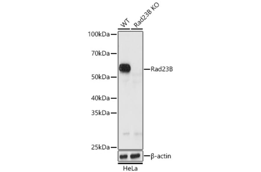
RAD23B expression in lysates from wild type (WT) and Rad23B knockout (KO) HeLa cells analyzed by western blot. Primary antibody incubation was performed for 1 hour on 25ug protein per lane with Anti-RAD23B Antibody (A12901) at a dilution of 1:1000 and detected with chemiluminescence.
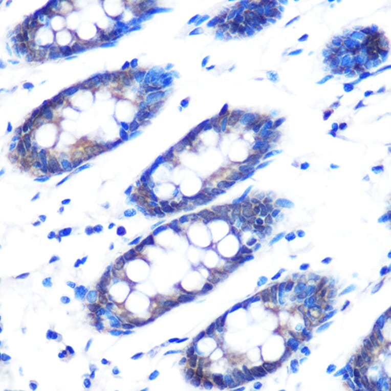
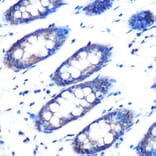
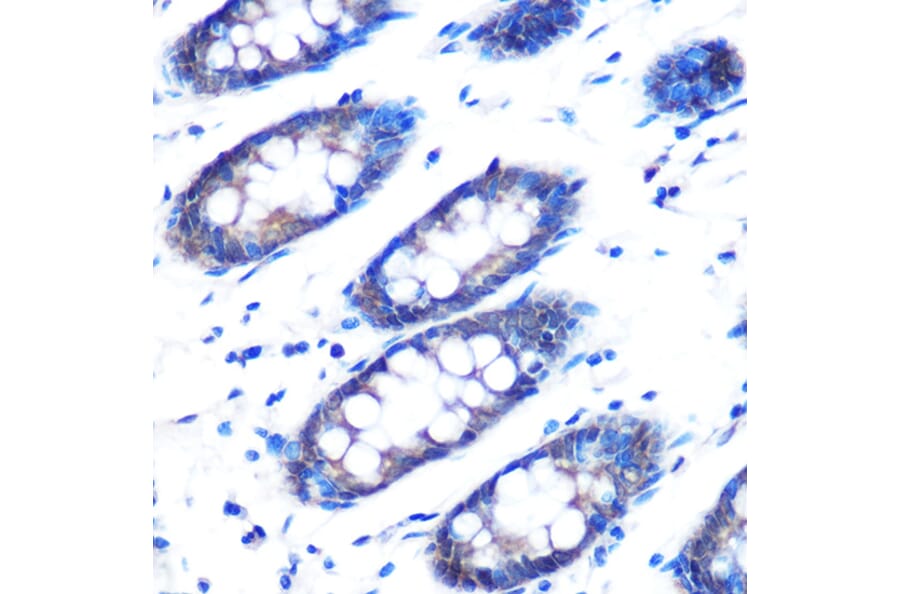
RAD23B expression in human colon tissue analyzed by immunohistochemistry. Tissue was paraffin-embedded, and antigen retrieval was achieved by microwaving in 10 mM PBS buffer, pH 7.2. Staining was performed with Anti-RAD23B Antibody (A12901) at a dilution of 1:100.
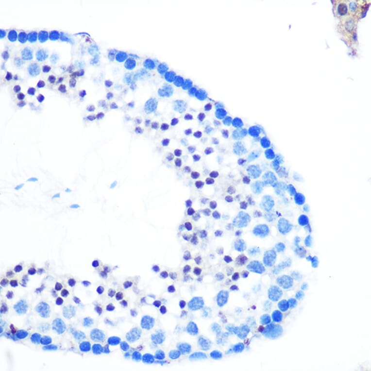
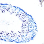
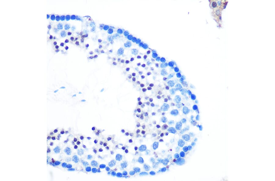
RAD23B expression in mouse testis tissue analyzed by immunohistochemistry. Tissue was paraffin-embedded, and antigen retrieval was achieved by microwaving in 10 mM PBS buffer, pH 7.2. Staining was performed with Anti-RAD23B Antibody (A12901) at a dilution of 1:100.
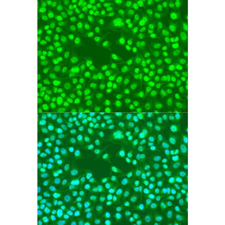
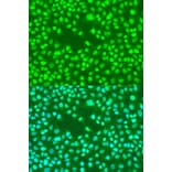
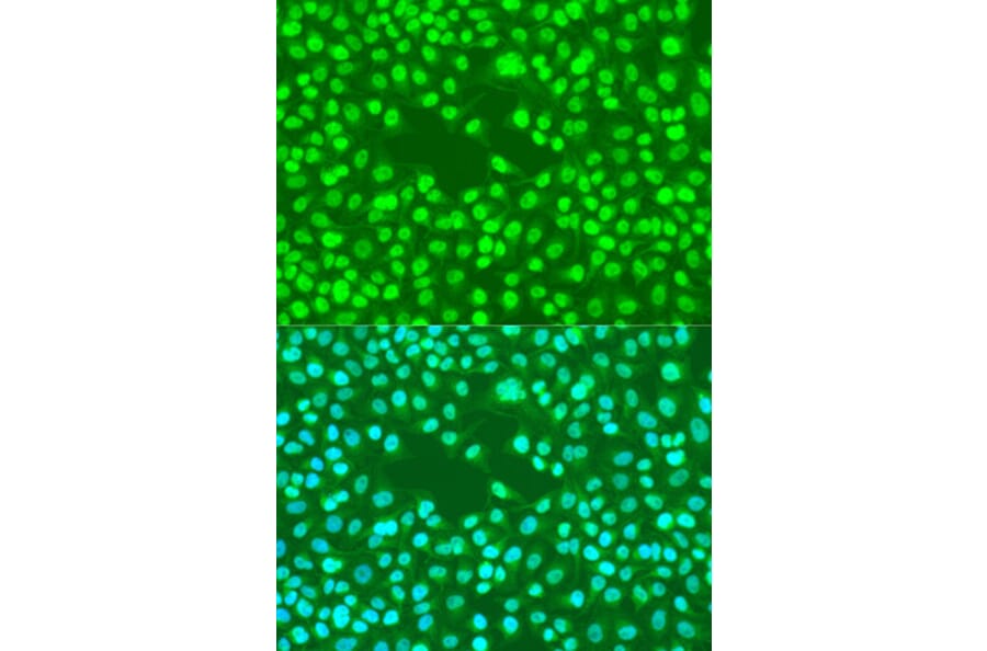
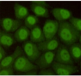
RAD23B expression in U2OS cells analyzed by immunofluorescence. Staining was performed with Anti-RAD23B Antibody (A12901) at a dilution of 1:100 followed by Cy3-conjugated Goat anti-Rabbit IgG (H+L) secondary antibody at a dilution of 1:500. Nuclei were stained with DAPI (blue).