Supplied in Phosphate Buffered Saline, pH 7.3, with 50% Glycerol and 0.05% Proclin 300.
Storage
Shipped at 4°C. Upon delivery aliquot and store at -20°C. Avoid freeze/thaw cycles.
Synonyms
ANX2LG, CAL1L, Calpactin I light chain, Calpactin-1 light chain, Cellular ligand of annexin II, CLP11, p10 protein, p11, Protein S100-A10, S100 calcium-binding protein A10
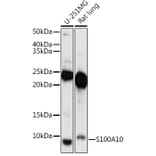
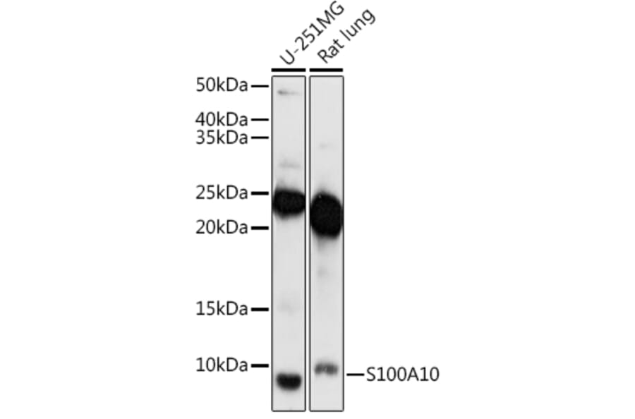
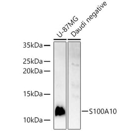
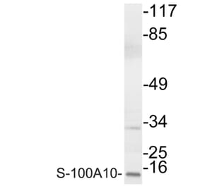
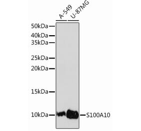
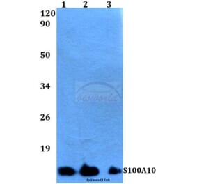
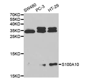
Western blot analysis of extracts of various cell lines, using Anti-S100A10 Antibody (A88015) at 1:1,000 dilution. The secondary antibody was Goat Anti-Rabbit IgG H&L Antibody (HRP) at 1:10,000 dilution. Lysates/proteins were present at 25µg per lane. The blocking buffer used was 3% non-fat dry milk in TBST. Detection was with a ECL Enhanced Kit. Exposure time: 180s.
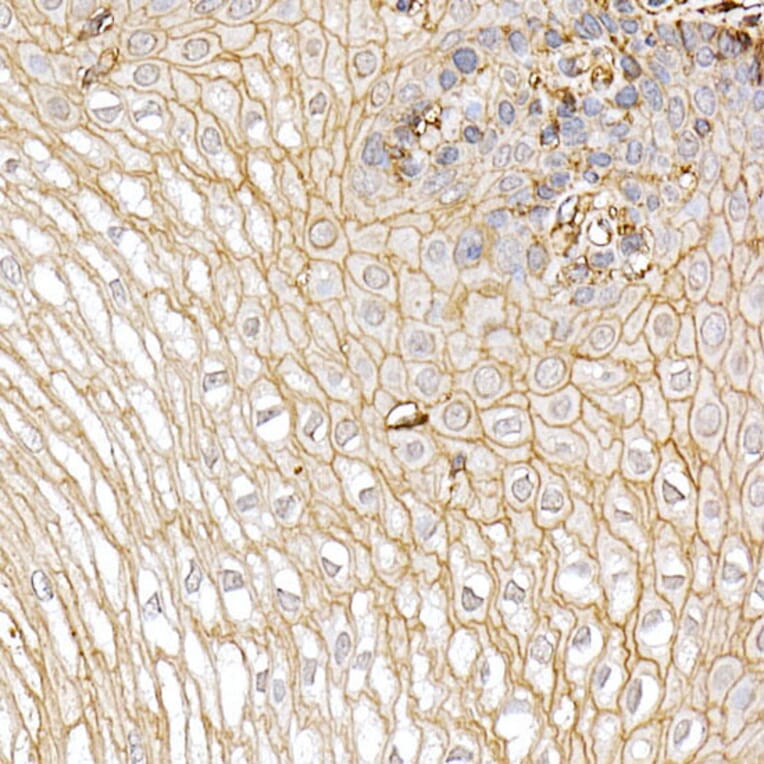
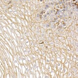
Immunohistochemistry analysis of paraffin-embedded human esophagus using Anti-S100A10 Antibody (A88015) at a dilution of 1:20 (40x lens). Perform high pressure antigen retrieval with 10 mM citrate buffer pH 6.0 before commencing with IHC staining protocol.
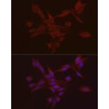
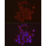
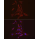
Immunofluorescence analysis of PC-12 using Anti-S100A10 Antibody (A88015) at a dilution of 1:20 (40x lens). DAPI was used to stain the cell nuclei (blue).
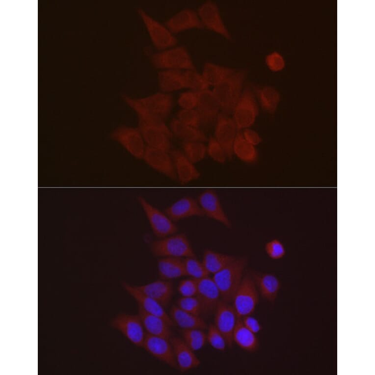
Immunofluorescence analysis of A-431 using Anti-S100A10 Antibody (A88015) at a dilution of 1:20 (40x lens). DAPI was used to stain the cell nuclei (blue).
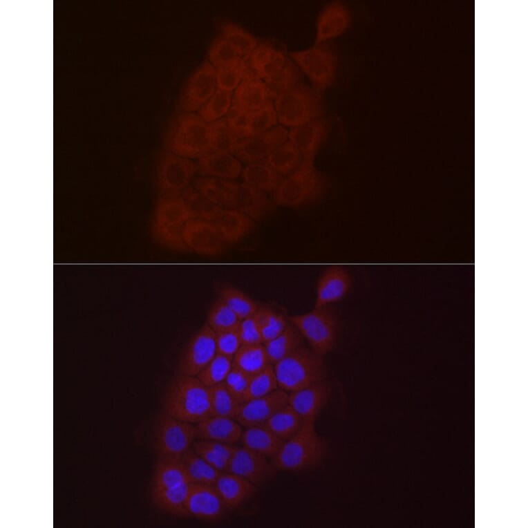
Immunofluorescence analysis of HeLa using Anti-S100A10 Antibody (A88015) at a dilution of 1:20 (40x lens). DAPI was used to stain the cell nuclei (blue).
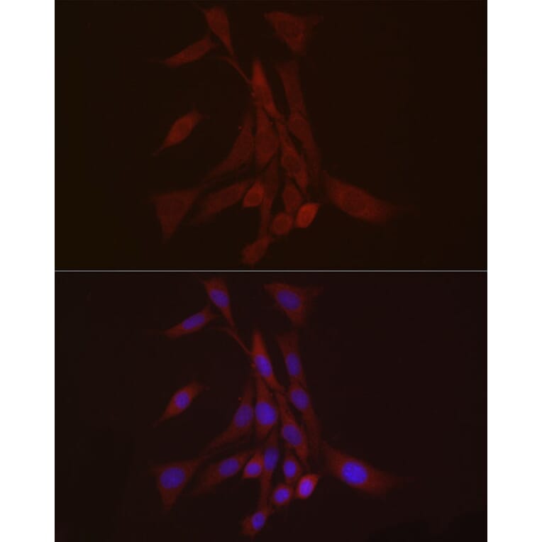
Immunofluorescence analysis of NIH/3T3 using Anti-S100A10 Antibody (A88015) at a dilution of 1:20 (40x lens). DAPI was used to stain the cell nuclei (blue).