This antibody binds specifically to the C-terminal domain of human S1P1, detecting the non-phosphorylated form. It is suitable for Western blot analysis, facilitates immunoprecipitation from tissue lysates, and is optimized for immunohistochemistry in cultured cells and tissue sections.
Applications
WB, IHC
Dilutions
WB: 1,000, IHC: 1:100
Reactivity
Human
Immunogen
Synthetic peptide corresponding to the C-terminal of human S1P1. Immunogen range is 22-22 amino acids.
Host
Rabbit
Clonality
Polyclonal
Isotype
IgG
Conjugate
Unconjugated
Purification
Antigen affinity purification.
Concentration
Lot Specific
Product Form
Liquid
Formulation
Supplied in Dulbecco's PBS, pH 7.4, with 150 mM NaCl and 0.005% Sodium Azide.
Storage
Shipped at 4°C. Upon delivery aliquot and store at -20°C. Avoid freeze/thaw cycles.
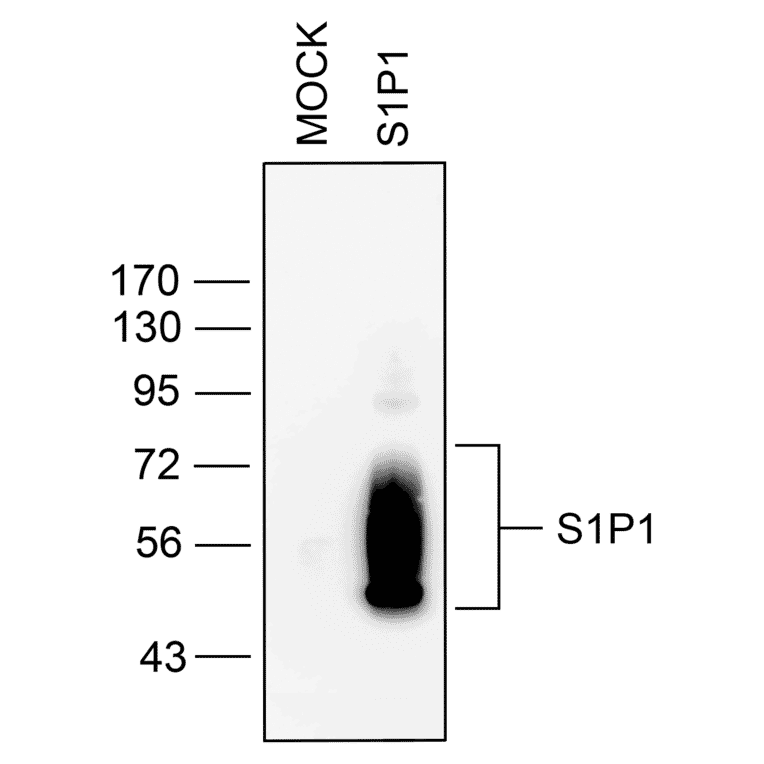
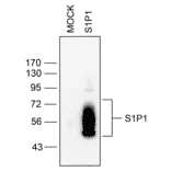
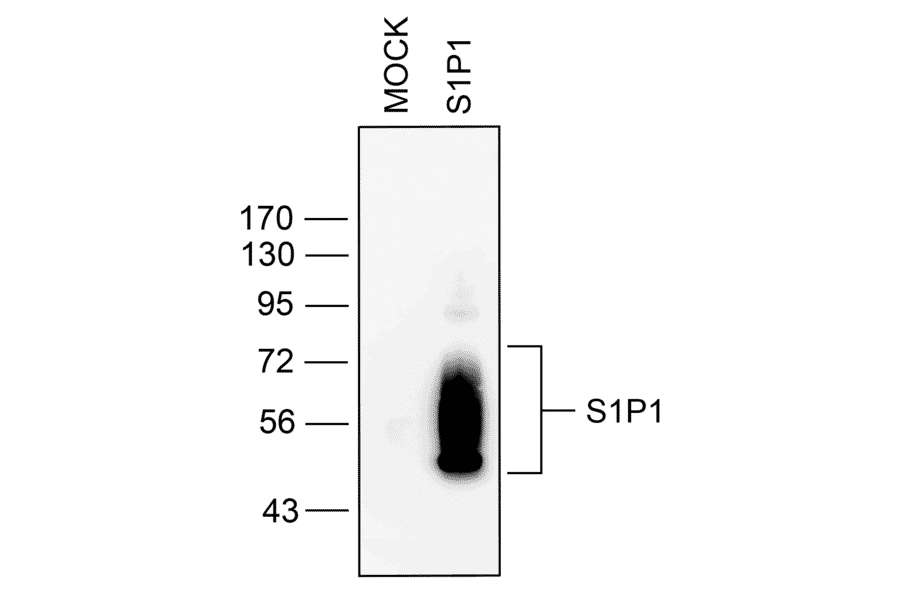
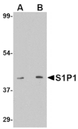
Western blot validation of S1P1 Receptor (S1P1) in transfected HEK293 cells. Lysates from native HEK293 cells (MOCK) or cells stably expressing S1P1 were probed with the phosphorylation-independent anti-S1P1 antibody (A334548) at a 1:100 dilution.
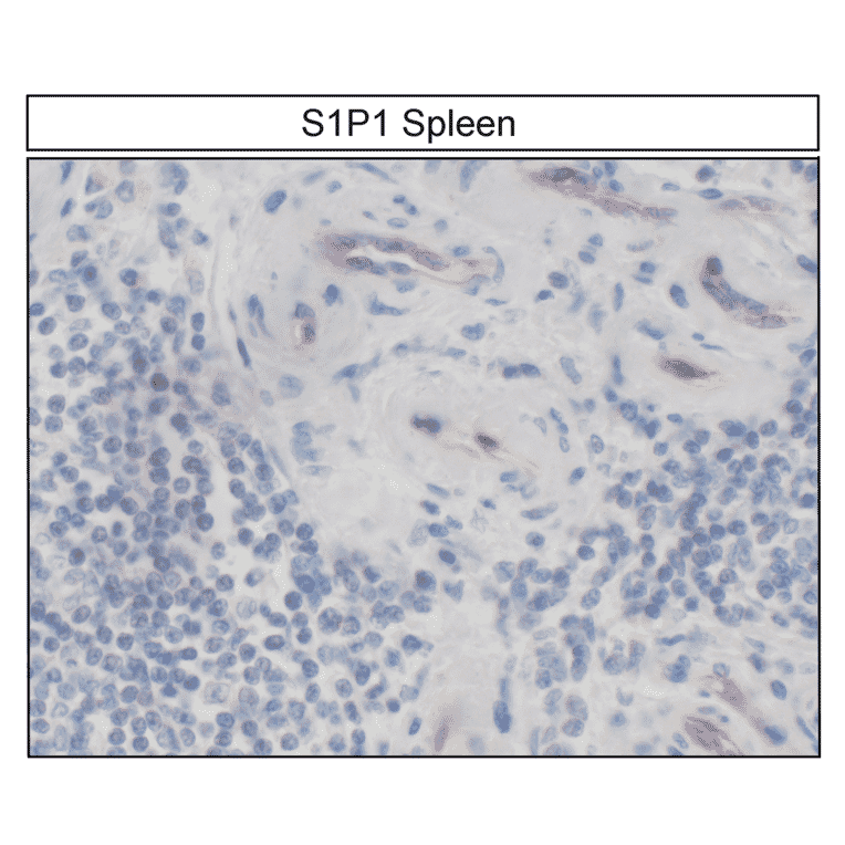
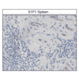
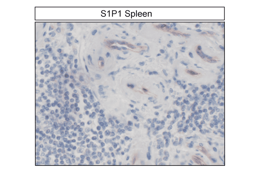
Localization of S1P1 Receptor (S1P1) in human spleen by immunohistochemistry. Spleen sections were dewaxed, microwaved in citric acid, and stained with anti-S1P1 antibody (A334548) at 1:100, followed by biotinylated anti-rabbit IgG and avidin-biotin solution. Color was developed with 3-amino-9-ethylcarbazole (AEC), and sections were counterstained with hematoxylin. S1P1 receptors were uniformly detected at the plasma membrane of nearly all spleen cells.
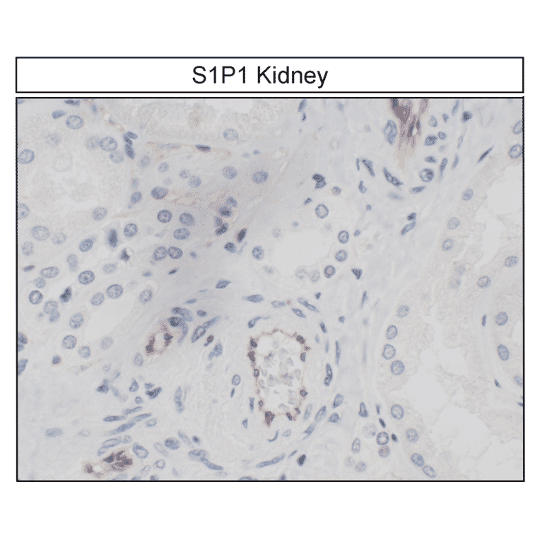
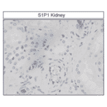
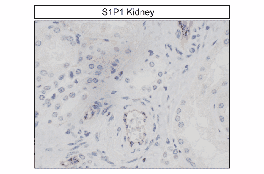
Detection of S1P1 Receptor (S1P1) in human kidney by immunohistochemistry. Kidney sections were dewaxed, microwaved in citric acid, and incubated with anti-S1P1 antibody (A334548) at 1:100, followed by biotinylated anti-rabbit IgG and avidin-biotin solution. Color was developed with 3-amino-9-ethylcarbazole (AEC), and sections were counterstained with hematoxylin. S1P1 receptors were observed at the plasma membrane of kidney cells.
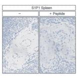
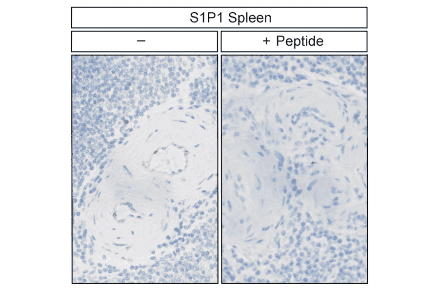
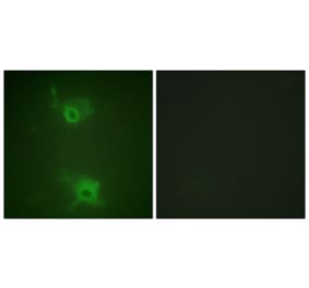
Validation of S1P1 Receptor (S1P1) antibody specificity in human spleen by immunohistochemistry. Spleen sections were dewaxed, microwaved in citric acid, and incubated without (left panel) or with (right panel) the peptide used to generate anti-S1P1 antibody (A334548). Sections were stained with anti-S1P1 antibody (A334548) at 1:100, followed by biotinylated anti-rabbit IgG and avidin-biotin solution, then developed with 3,3-diaminobenzidine (DAB)-glucose oxidase and lightly counterstained with hematoxylin. S1P1 receptors were detected at the plasma membrane only in sections without peptide incubation.
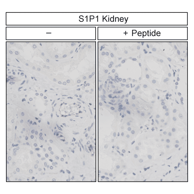
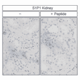
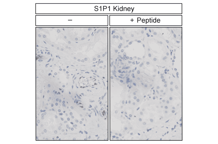
Confirmation of S1P1 Receptor (S1P1) antibody specificity in human kidney by immunohistochemistry. Kidney sections were dewaxed, microwaved in citric acid, and incubated without (left panel) or with (right panel) the peptide used to generate anti-S1P1 antibody (A334548). Sections were stained with anti-S1P1 antibody (A334548) at 1:100, followed by biotinylated anti-rabbit IgG and avidin-biotin solution, then developed with 3,3-diaminobenzidine (DAB)-glucose oxidase and lightly counterstained with hematoxylin. S1P1 receptors were observed at the plasma membrane only in sections without peptide incubation.
Recombinant humanized monoclonal antibody to S1P1 for use as a research grade Sonepcizumab biosimila for ELISA, Flow Cytometry, Functional Studies and in vivo Research.
Recombinant humanized monoclonal antibody to S1P1 for use as a research grade Sonepcizumab biosimila for ELISA, Flow Cytometry, Functional Studies and in vivo Research.