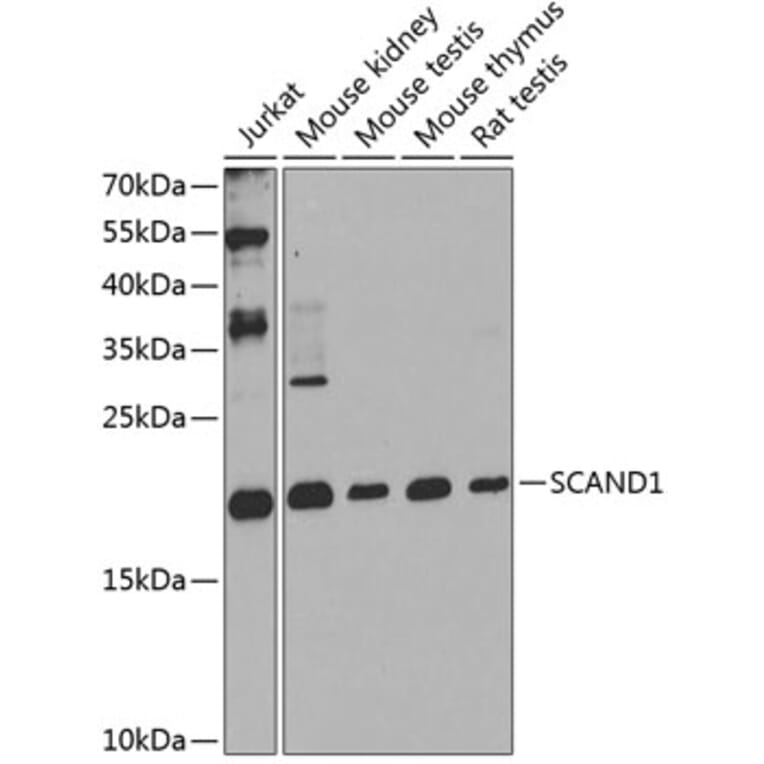
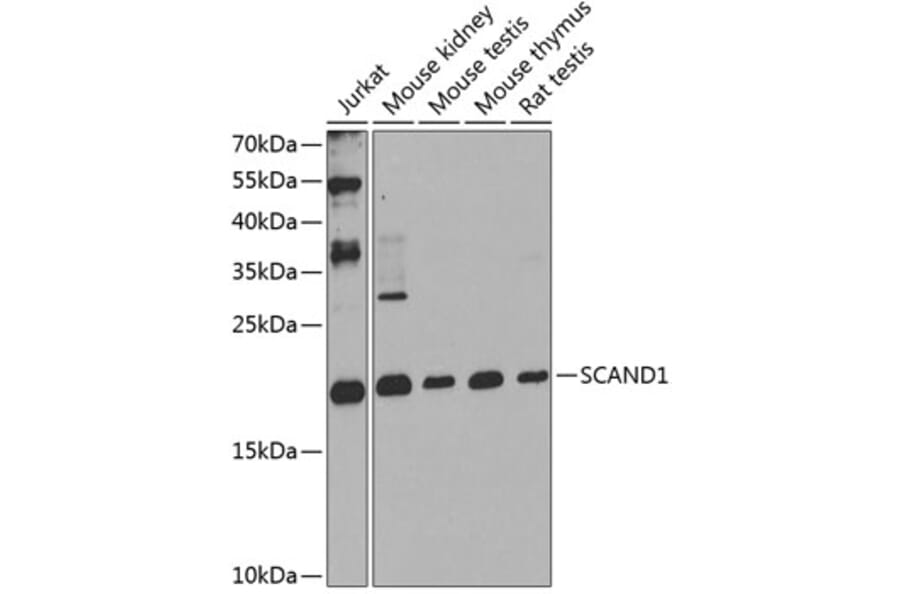
Western blot analysis of extracts of various cell lines, using Anti-SCAND1 Antibody (A16153) at 1:1,000 dilution. The secondary antibody was Goat Anti-Rabbit IgG H&L Antibody (HRP) at 1:10,000 dilution. Lysates/proteins were present at 25µg per lane. The blocking buffer used was 3% non-fat dry milk in TBST. Detection was with a ECL Enhanced Kit. Exposure time: 90s.
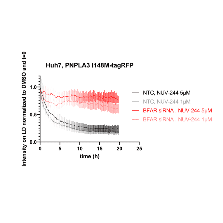
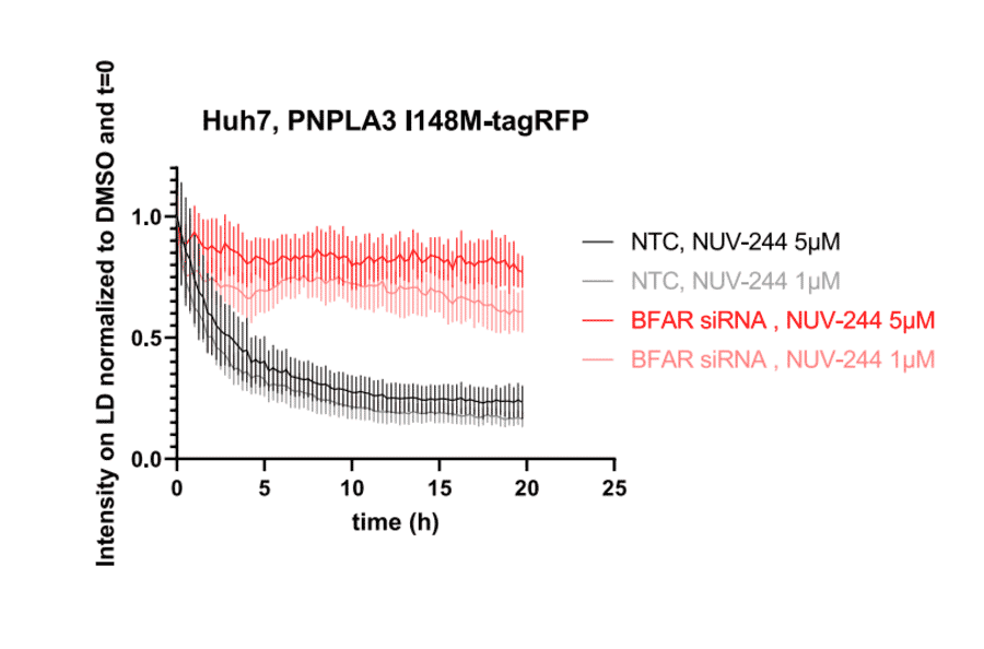
Quantitative live-cell fluorescence analysis of PNPLA3 I148M-tagRFP intensity on lipid droplets in Huh7 cells treated with non-targeting control siRNA or BFAR siRNA and exposed to 1 µM or 5 µM NUV-244. Data are normalized to DMSO control at t = 0 h. BFAR knockdown partially rescues PNPLA3 I148M-tagRFP from NUV-244-induced degradation. Values represent mean ± SD (n = 9 per condition). Two independent experiments with comparable results are shown.
Figure 5D from Steigemann P et al. (2025). Originally published in iScience, DOI: 10.1016/j.isci.2025.112384.. Reproduced under CC BY 4.0.
Publishing research using Anti-SCAND1 Antibody (A16153)? Please let us know so that we can list the citation on this page.
Alternative products to Anti-SCAND1 Antibody (A16153)