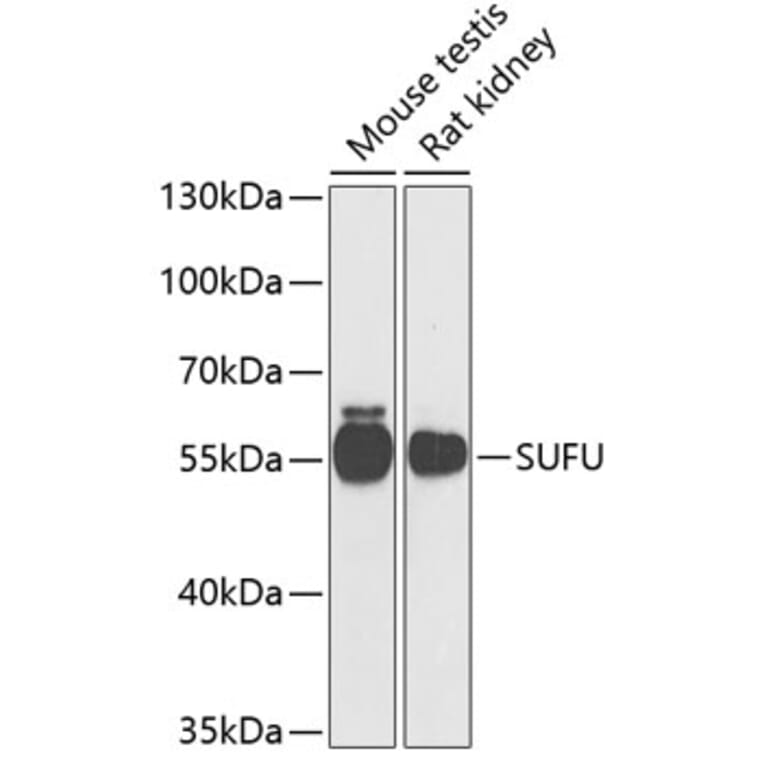
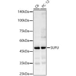
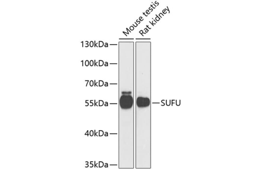
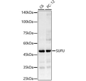
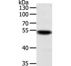
SUFU expression in various lysates analyzed by western blot. Primary antibody incubation was performed for 1 hour on 25ug protein per lane with Anti-SUFU Antibody (A15365) at a dilution of 1:1000 and detected with chemiluminescence.
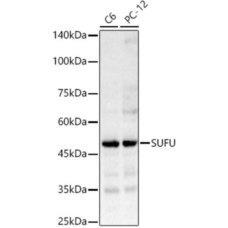
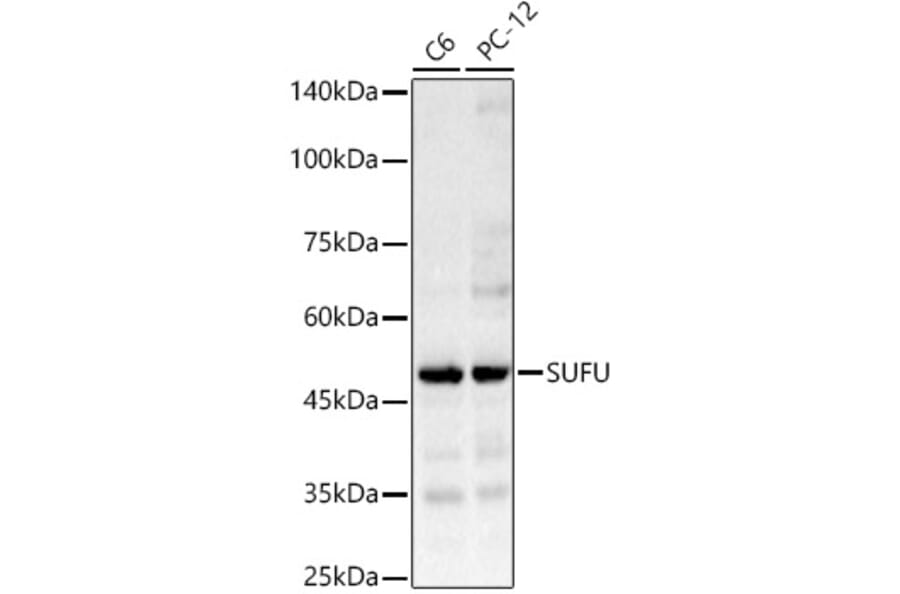
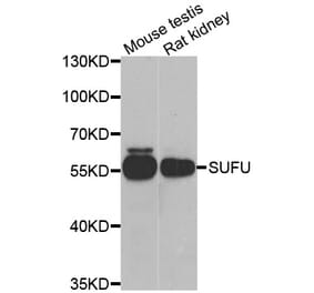
SUFU expression in various lysates analyzed by western blot. Primary antibody incubation was performed for 1 hour on 25ug protein per lane with Anti-SUFU Antibody (A15365) at a dilution of 1:1000 and detected with chemiluminescence.
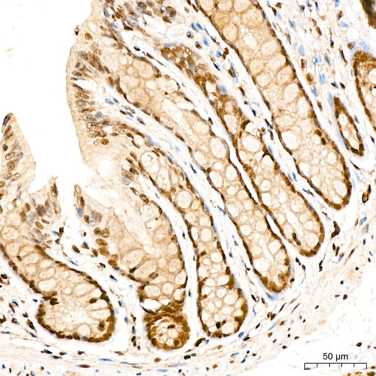
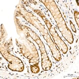
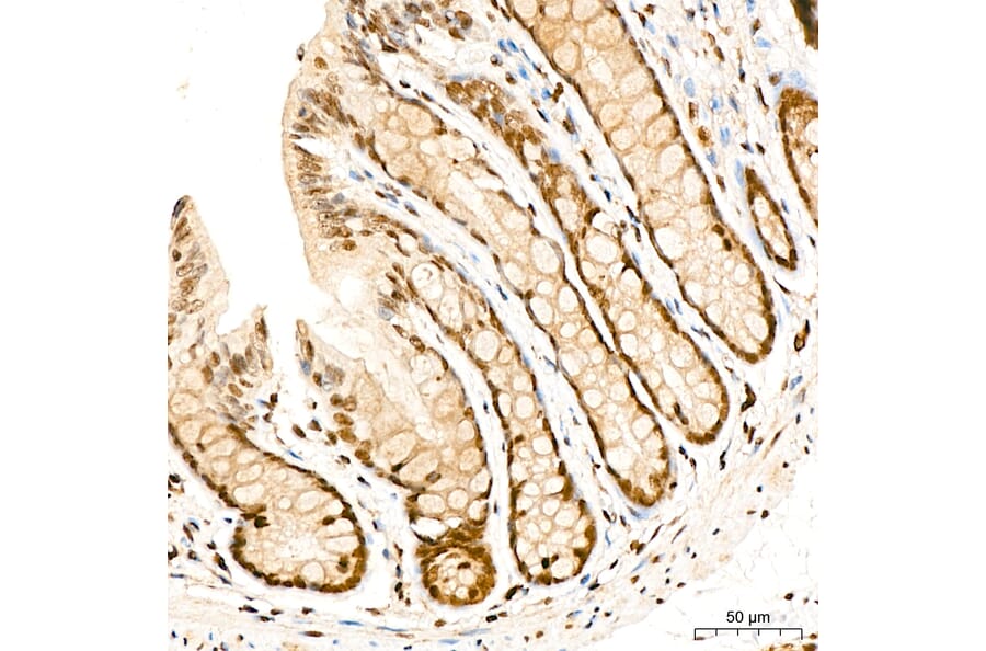
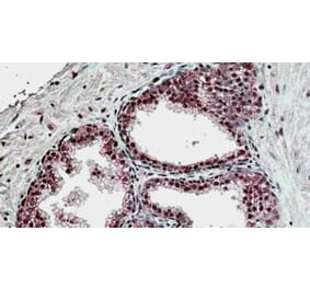
SUFU expression in mouse colon tissue analyzed by immunohistochemistry. Tissue was paraffin-embedded, and antigen retrieval was achieved with 10 mM citrate buffer, pH 6.0, under high pressure. Staining was performed with Anti-SUFU Antibody (A15365) at a dilution of 1:400.
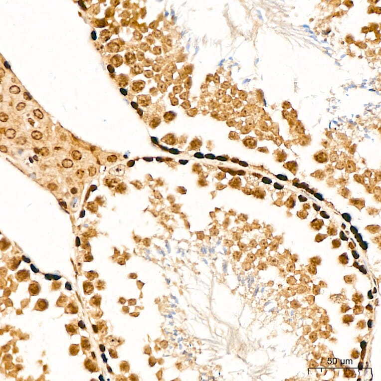
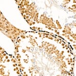
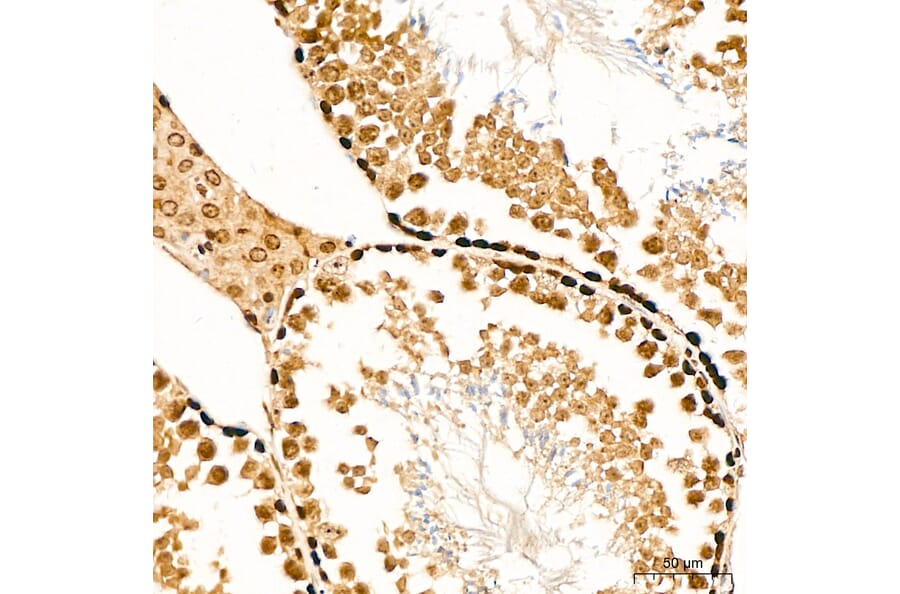
SUFU expression in mouse testis tissue analyzed by immunohistochemistry. Tissue was paraffin-embedded, and antigen retrieval was achieved with 10 mM citrate buffer, pH 6.0, under high pressure. Staining was performed with Anti-SUFU Antibody (A15365) at a dilution of 1:400.
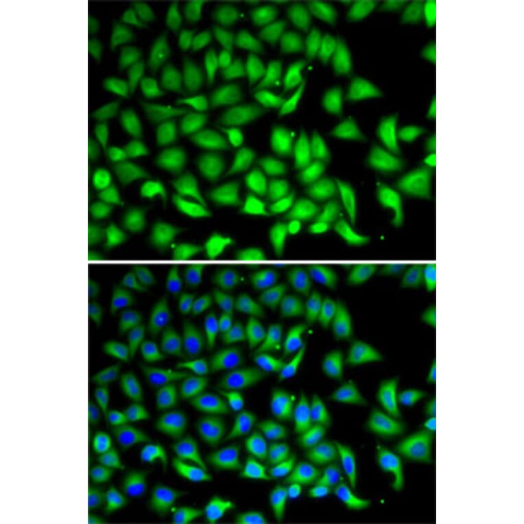
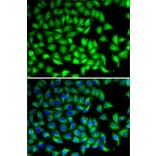
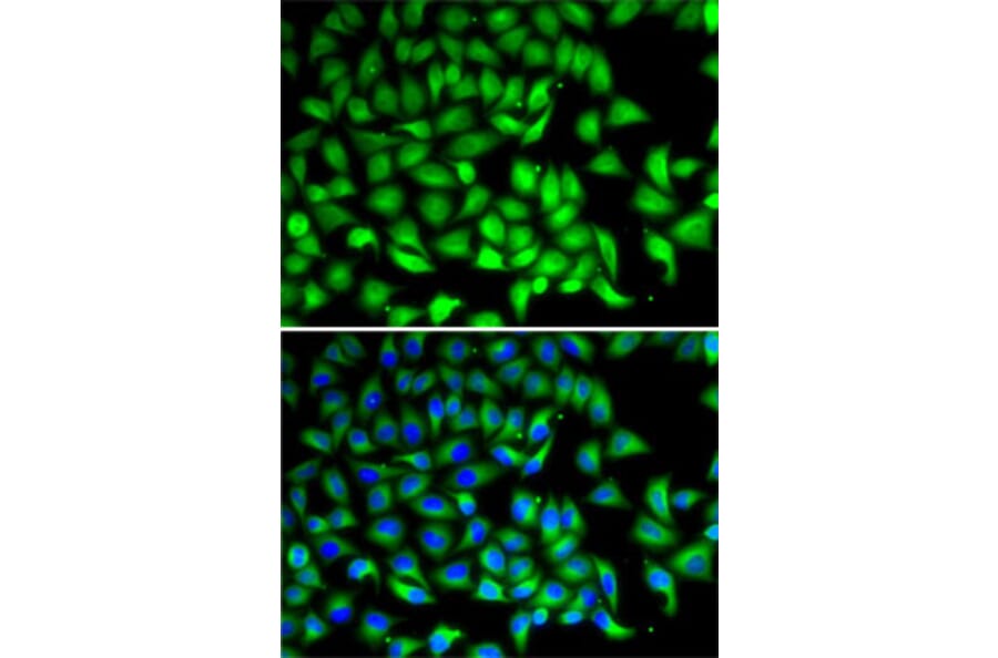
SUFU expression in MCF7 cells analyzed by immunofluorescence. Staining was performed with Anti-SUFU Antibody (A15365) followed by Cy3 Goat Anti-Rabbit IgG (H+L) secondary antibody at a dilution of 1:500. Nuclei were stained with DAPI (blue).