The epitope for this antibody is in the center of the peptide HNSNRQLERSGRFGGNPGGF, C-terminal to second RRM domain, amino acids 264-283 of human TDP43. This is the region typically in TDP43 inclusions. This peptide is 100% conserved in a wide variety of mammalian species.
Applications
WB, ICC/IF, IHC
Dilutions
WB: 1:5,000, ICC/IF: 1:1,000, IHC: 1:1,000
Reactivity
Human, Rat, Mouse, Bovine, Porcine, Horse
Immunogen
Recombinant full-length human TDP43, expressed in and purified from E. coli.
Host
Mouse
Clonality
Monoclonal
Clone ID
3H8
Isotype
IgG1
Light Chains
kappa
Conjugate
Unconjugated
Purification
Immunogen affinity purification.
Concentration
1 mg/ml
Molecular Weight
43 kDa
Product Form
Liquid
Formulation
Supplied in Phosphate Buffered Saline with 50% Glycerol and 5mM Sodium Azide.
Storage
Shipped at 4°C. Upon delivery aliquot and store at -20°C. Avoid freeze/thaw cycles.
Immunofluorescent analysis of rat hippocampus section stained with Anti-TARBDP Antibody, at a dilution of 1:2,000, in red, and Anti-GFAP Antibody (A85307 | 1:5,000, in green. The blue is DAPI staining of nuclear DNA. Following transcardial perfusion of rat with 4% paraformaldehyde, brain was post fixed for 24 hours, cut to 45 µM, and free-floating sections were stained with the above antibodies. The TDP43 protein is concentrated in the nuclei of hippocampal neurons, while the Anti-GFAP Antibody stains the network of astroglial cells.
Anti-TARBDP Antibody was used to stain a section of formalin fixed adult rat brain, specifically the hippocampus. Hippocampal neuron nuclei are stained strongly. Anti-GFAP Antibody (A85307 | green) shows the processes of astrocytic glial cells. Nuclei of all cells are revealed with DAPI DNA stain (blue). The Anti-TARDP Antibody stains neuronal nuclei strongly and the nuclei of some non-neuronal cells much more weakly. Neuronal nuclei therefore look crimson, since they are both red due to the content of TDP43 and blue due to their content of DNA, stained blue with DAPI.
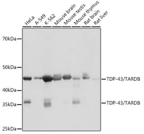
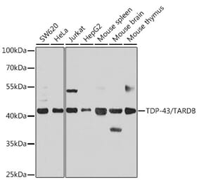
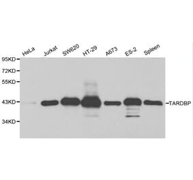
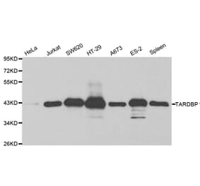
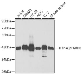
Western blot analysis of whole brain lysates and nuclear extract from whole brain using Anti-TARBDP Antibody [3H8] (A85389), at a dilution of 1:2,000, in green. The lanes contain samples of: [Lane 1] Protein standards, in red, [Lane 2] rat brain, [Lane 3] rat brain nuclear extract, [Lane 4] mouse brain, [Lane 5] mouse brain nuclear extract. The strong band at 43 kDa visible in both whole brain and brain nuclear extract corresponds to TARBDP protein.
Immunohistochemistry analysis of a 4% PFA fixed paraffin embedded rat cerebellum section with Anti-TDP43 Antibody [3H8] (A85389) at a dilution of 1:4,000 detected with DAB (brown) using the Vector Labs ImmPRESS method and reagents with citra buffer retrieval. Counterstained with Hematoxylin (blue). The Anti-TDP43 Antibody [3H8] (A85389) strongly and specifically labels nuclei. Note: this antibody performs well in testing with both 4% PFA and standard NBF fixed tissues.
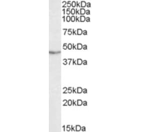
Western blot analysis of crude extract of mouse brain nuclear fraction (left lane) and cytoplasmic fraction (right lane) probed with Anti-TARBDP Antibody. There is a strong clear band in the nuclear preparation running at 43 kDa, and a much more minor band in the cytoplasmic fraction.
Immunohistochemistry analysis of ethanol fixed paraffin embedded human brain sections from a patient with fronto-temporal dementia using Anti-TDP43 Antibody [3H8] (A85389). The antibody is a robust marker of TDP43 positive inclusions.


![Immunofluorescence - Anti-TDP43 Antibody [3H8] (A85389) - Antibodies.com](https://cdn.antibodies.com/image/catalog/85/A85389_1.jpg?profile=product_top)
![Immunofluorescence - Anti-TDP43 Antibody [3H8] (A85389) - Antibodies.com](https://cdn.antibodies.com/image/catalog/85/A85389_2.jpg?profile=product_top)
![Western Blot - Anti-TDP43 Antibody [3H8] (A85389) - Antibodies.com](https://cdn.antibodies.com/image/catalog/85/A85389_3.jpg?profile=product_top)
![Immunohistochemistry - Anti-TDP43 Antibody [3H8] (A85389) - Antibodies.com](https://cdn.antibodies.com/image/catalog/85/A85389_4.jpg?profile=product_top)
![Western Blot - Anti-TDP43 Antibody [3H8] (A85389) - Antibodies.com](https://cdn.antibodies.com/image/catalog/85/A85389_5.jpg?profile=product_top)
![Immunohistochemistry - Anti-TDP43 Antibody [3H8] (A85389) - Antibodies.com](https://cdn.antibodies.com/image/catalog/85/A85389_6.jpg?profile=product_top)
![Immunofluorescence - Anti-TDP43 Antibody [3H8] (A85389) - Antibodies.com](https://cdn.antibodies.com/image/catalog/85/A85389_1.jpg?profile=product_top_thumb)
![Immunofluorescence - Anti-TDP43 Antibody [3H8] (A85389) - Antibodies.com](https://cdn.antibodies.com/image/catalog/85/A85389_2.jpg?profile=product_top_thumb)
![Western Blot - Anti-TDP43 Antibody [3H8] (A85389) - Antibodies.com](https://cdn.antibodies.com/image/catalog/85/A85389_3.jpg?profile=product_top_thumb)
![Immunohistochemistry - Anti-TDP43 Antibody [3H8] (A85389) - Antibodies.com](https://cdn.antibodies.com/image/catalog/85/A85389_4.jpg?profile=product_top_thumb)
![Western Blot - Anti-TDP43 Antibody [3H8] (A85389) - Antibodies.com](https://cdn.antibodies.com/image/catalog/85/A85389_5.jpg?profile=product_top_thumb)
![Immunohistochemistry - Anti-TDP43 Antibody [3H8] (A85389) - Antibodies.com](https://cdn.antibodies.com/image/catalog/85/A85389_6.jpg?profile=product_top_thumb)
![Immunofluorescence - Anti-TDP43 Antibody [3H8] (A85389) - Antibodies.com](https://cdn.antibodies.com/image/catalog/85/A85389_1.jpg?profile=product_image)
![Immunofluorescence - Anti-TDP43 Antibody [3H8] (A85389) - Antibodies.com](https://cdn.antibodies.com/image/catalog/85/A85389_2.jpg?profile=product_image)
![Western Blot - Anti-TDP43 Antibody [3H8] (A85389) - Antibodies.com](https://cdn.antibodies.com/image/catalog/85/A85389_3.jpg?profile=product_image)
![Immunohistochemistry - Anti-TDP43 Antibody [3H8] (A85389) - Antibodies.com](https://cdn.antibodies.com/image/catalog/85/A85389_4.jpg?profile=product_image)
![Western Blot - Anti-TDP43 Antibody [3H8] (A85389) - Antibodies.com](https://cdn.antibodies.com/image/catalog/85/A85389_5.jpg?profile=product_image)
![Immunohistochemistry - Anti-TDP43 Antibody [3H8] (A85389) - Antibodies.com](https://cdn.antibodies.com/image/catalog/85/A85389_6.jpg?profile=product_image)