Synthetic peptide corresponding to the C terminal region of mouse TORC2.
Sequence
C-DPAVEDSFRSDRLQ
Host
Goat
Clonality
Polyclonal
Isotype
IgG
Conjugate
Unconjugated
Purification
Purified from goat serum by ammonium sulphate precipitation followed by antigen affinity chromatography using the immunizing peptide.
Concentration
500 µg/ml
Molecular Weight
90 kDa
Predicted MW
73.2kDa
Product Form
Liquid
Formulation
Supplied in Tris Buffered Saline, pH 7.3, with 0.5% BSA and 0.02% Sodium Azide.
Storage
Shipped at 4°C. Upon delivery aliquot and store at -20°C. Avoid freeze/thaw cycles.
Synonyms
CREB-regulated transcription coactivator 2, CRTC2, TORC-2, Transducer of CREB protein 2, Transducer of regulated cAMP response element-binding protein 2
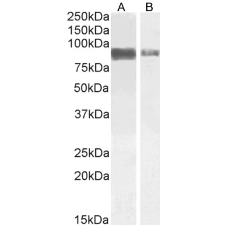
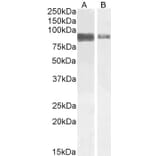
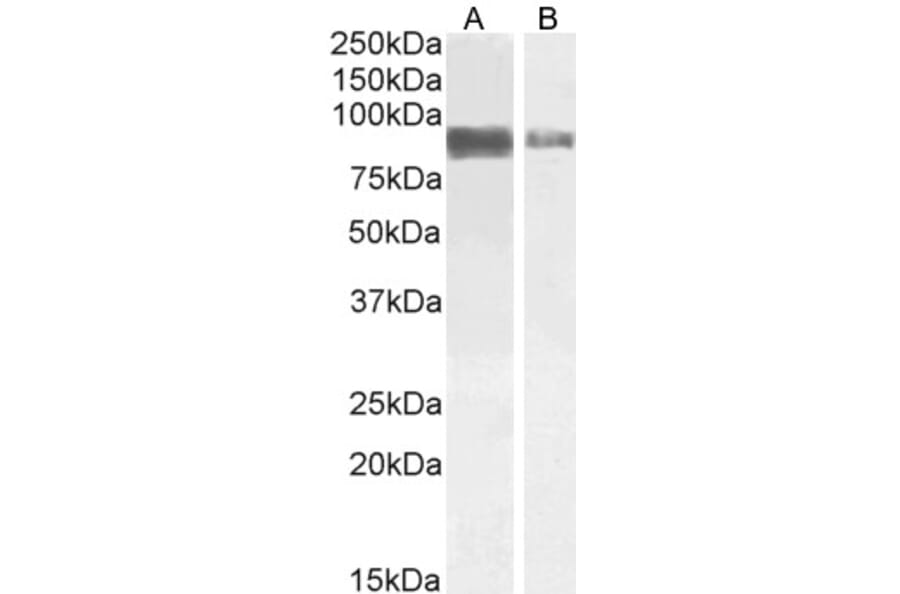
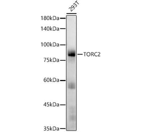
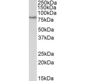
TORC2 expression in Jurkat (A) and A431 (B) cell lysate analyzed by western blot. Cells were lysed in RIPA buffer and 35µg protein was run per lane. Primary incubation was performed with Anti-TORC2 Antibody (A84934) at 1µg/ml (A) or 2µg/ml (B) and detected by chemiluminescence.
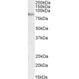
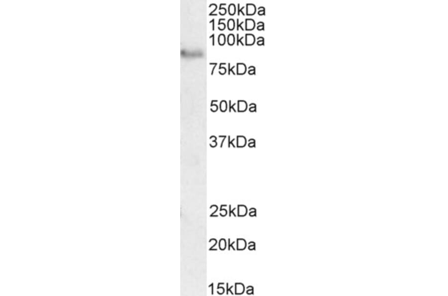
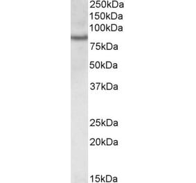
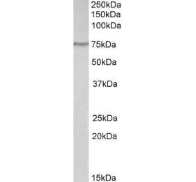
TORC2 expression in Rat Brain lysate analyzed by western blot. Cells were lysed in RIPA buffer and 35µg protein was run per lane. Primary antibody incubation was performed with Anti-TORC2 Antibody (A84934) at 2µg/ml and detected by chemiluminescence.
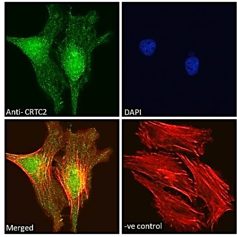
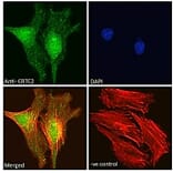
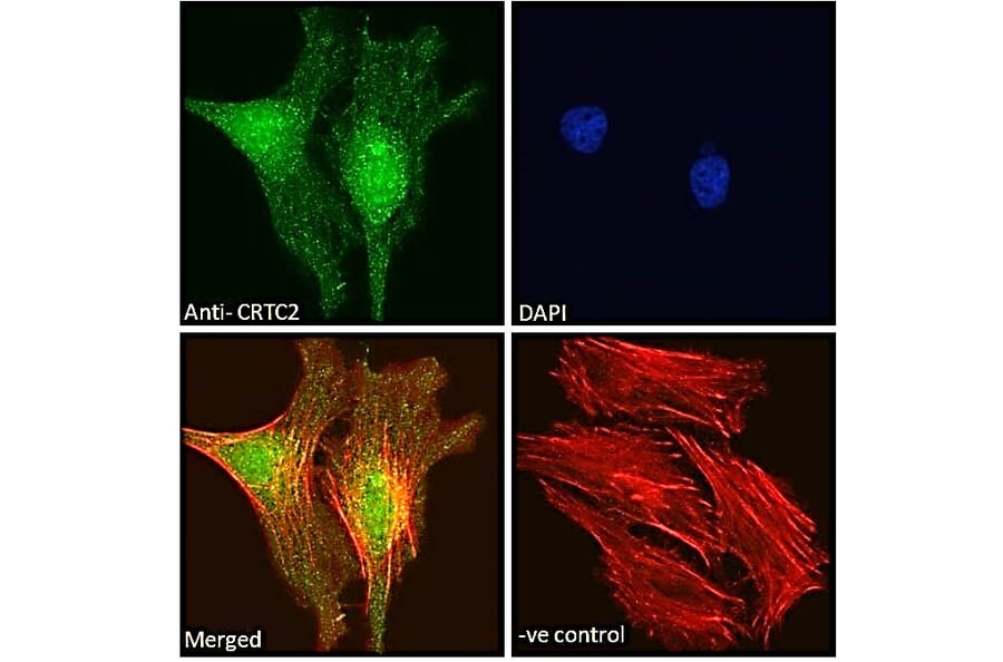
TORC2 expression in HeLa cells analyzed by immunofluorescence. Cells were permeabilized with 0.15% Triton. Staining was performed with Anti-TORC2 Antibody (A84934) at 10µg/ml for 1 hour and Alexa Fluor 488 secondary antibody at 2µg/ml. Strong nuclear staining shown and nuclei were stained with DAPI (blue) while actin filaments were stained with phalloidin (red). Negative control: Goat IgG Isotype Control at 10µg/ml followed by Alexa Fluor 488 secondary antibody at 2µg/ml.
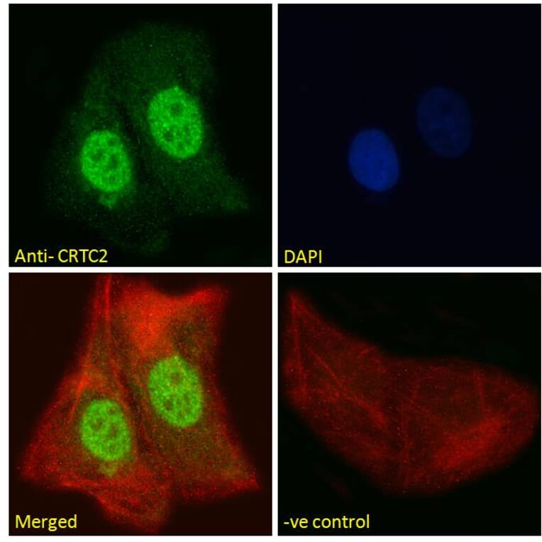
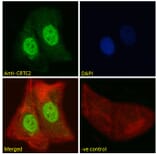
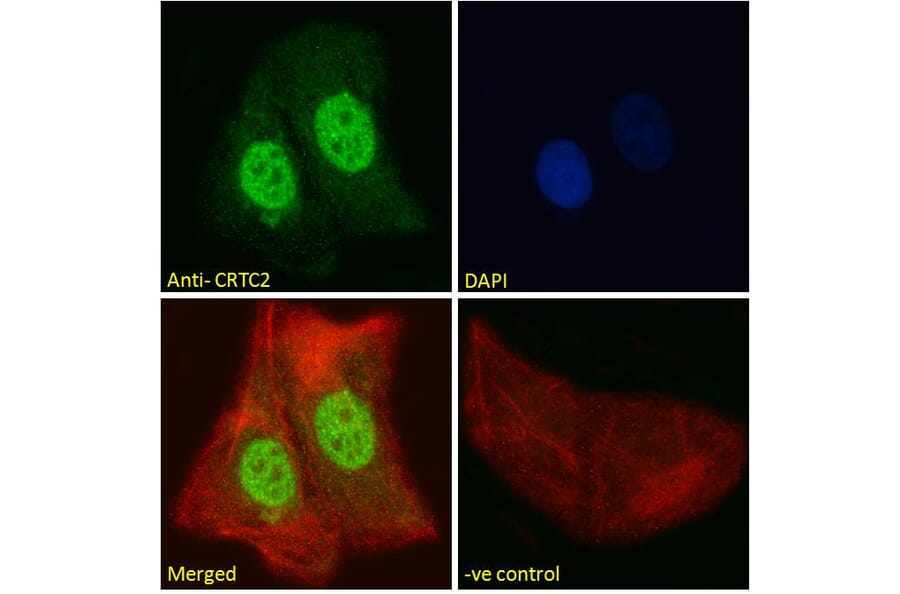
TORC2 expression in U2OS cells analyzed by immunofluorescence. Cells were permeabilized with 0.15% Triton. Staining was performed with Anti-TORC2 Antibody (A84934) at 10µg/ml for 1 hour and Alexa Fluor 488 secondary antibody at 2µg/ml. Strong nuclear staining shown and nuclei were stained with DAPI (blue) while actin filaments were stained with phalloidin (red). Negative control: Goat IgG Isotype Control at 10µg/ml followed by Alexa Fluor 488 secondary antibody at 2µg/ml.
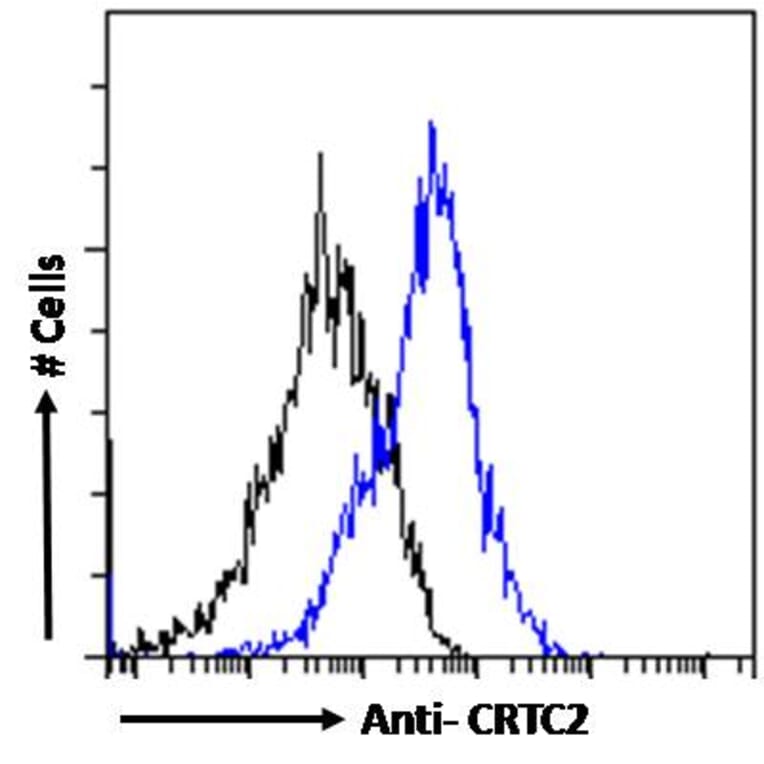
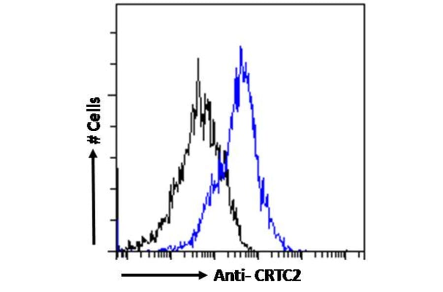
TORC2 expression in HeLa cells (blue line) analyzed by flow cytometry. Cells were fixed in PFA and permeabilized with 0.5% Triton. Staining was performed with Anti-TORC2 Antibody (A84934) at 10µg/ml for 1 hour and Alexa Fluor 488 secondary antibody at 1µg/ml. Negative Control: Goat IgG Isotype Control (black line) followed by Alexa Fluor 488 secondary antibody.