Supplied in Phosphate Buffered Saline, pH 7.3, with 50% Glycerol and 0.01% Thiomersal.
Storage
Shipped at 4°C. Upon delivery aliquot and store at -20°C. Avoid freeze/thaw cycles.
Synonyms
Heat shock protein 75 kDa, mitochondrial, Heat shock protein family C member 5, HSP 75, HSP75, HSPC5, TNFR-associated protein 1, TRAP-1, Tumor necrosis factor type 1 receptor-associated protein
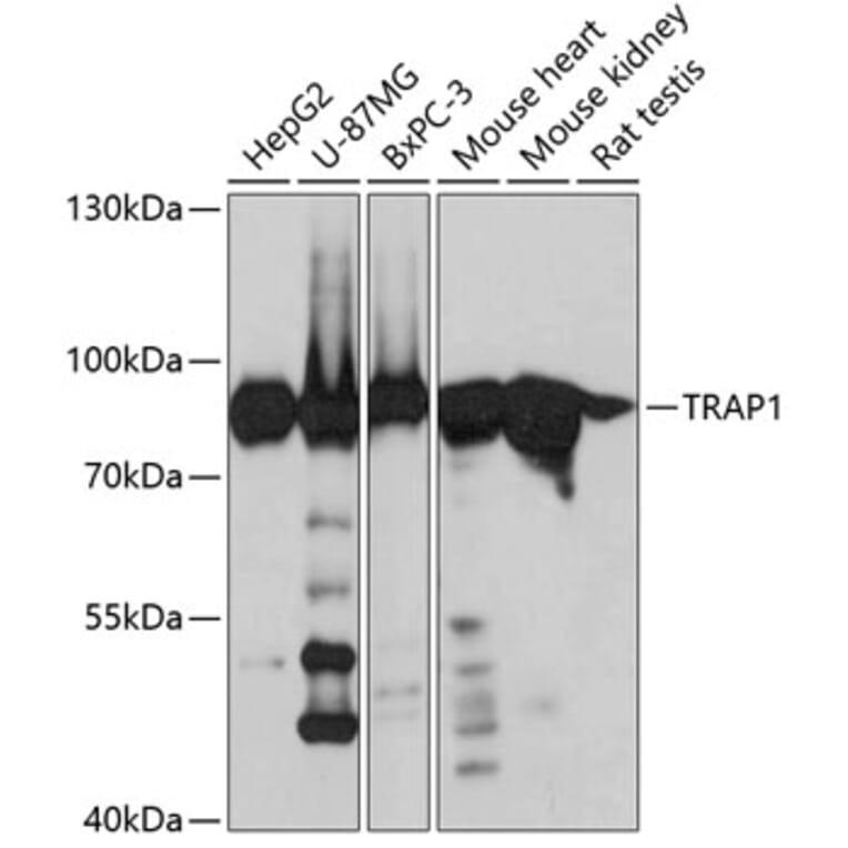
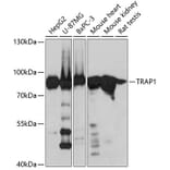
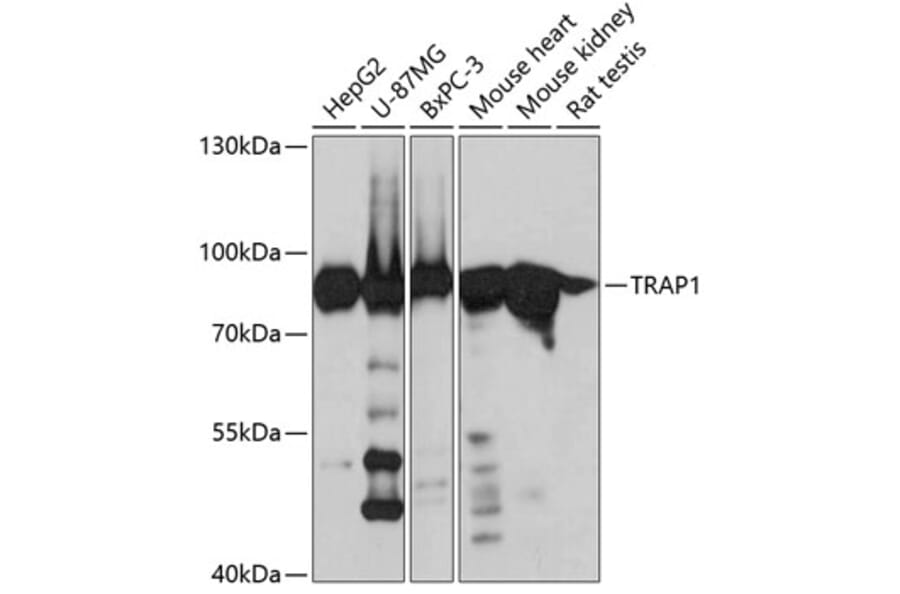
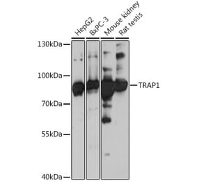
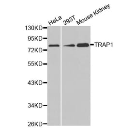
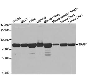
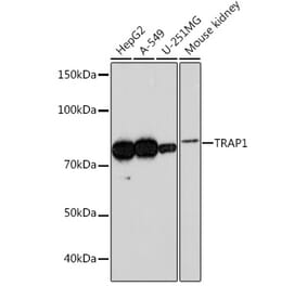
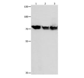
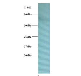
Western blot analysis of extracts of various cell lines, using Anti-TRAP1 Antibody (A14167) at 1:3,000 dilution. The secondary antibody was Goat Anti-Rabbit IgG H&L Antibody (HRP) at 1:10,000 dilution. Lysates/proteins were present at 25µg per lane. The blocking buffer used was 3% non-fat dry milk in TBST. Detection was with a ECL Basic Kit. Exposure time: 30s.
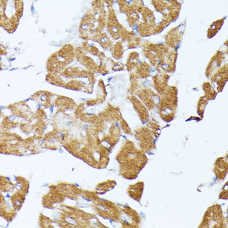
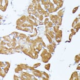
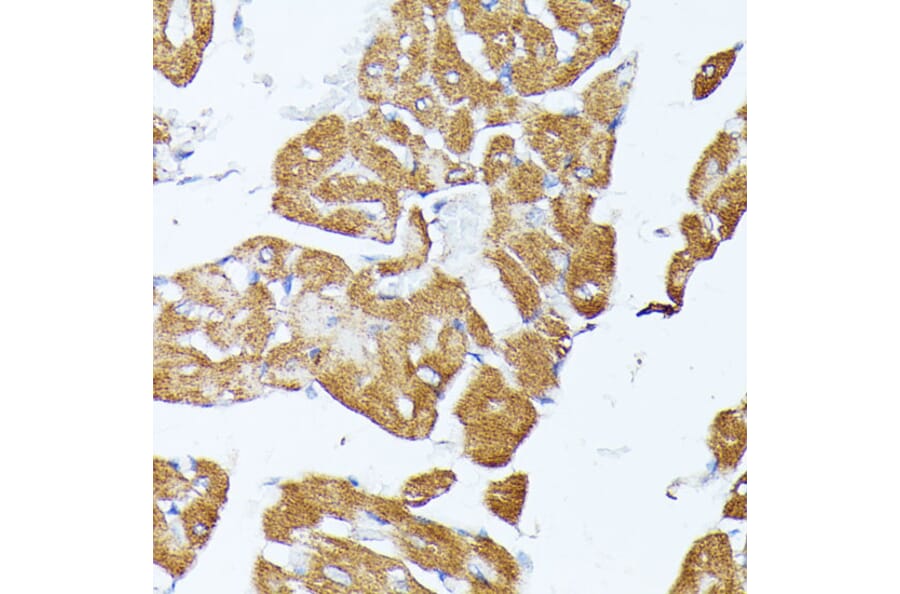
Immunohistochemistry analysis of paraffin-embedded rat heart using Anti-TRAP1 Antibody (A14167) at a dilution of 1:100 (40x lens). Perform microwave antigen retrieval with 10 mM PBS buffer pH 7.2 before commencing with IHC staining protocol.
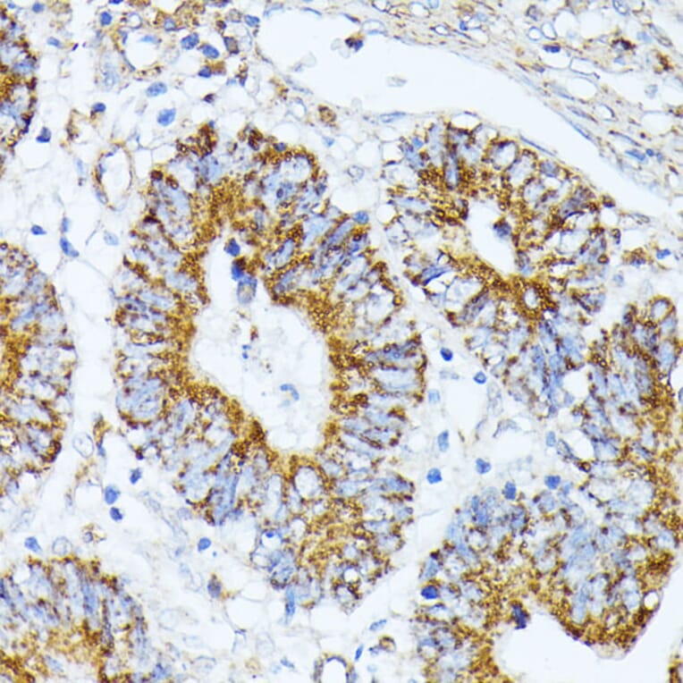
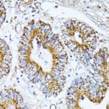
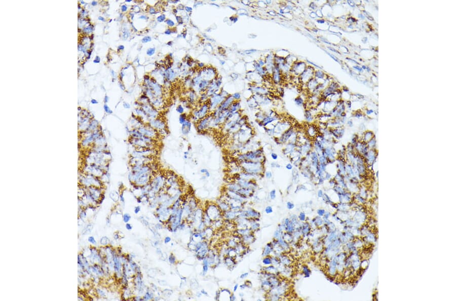
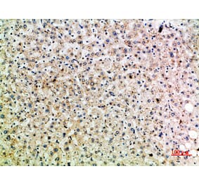
Immunohistochemistry analysis of paraffin-embedded human colon carcinoma tissue using Anti-TRAP1 Antibody (A14167) at a dilution of 1:100 (40x lens). Perform microwave antigen retrieval with 10 mM PBS buffer pH 7.2 before commencing with IHC staining protocol.
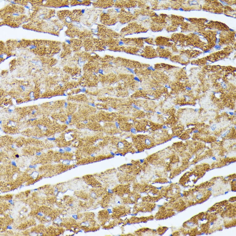
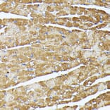
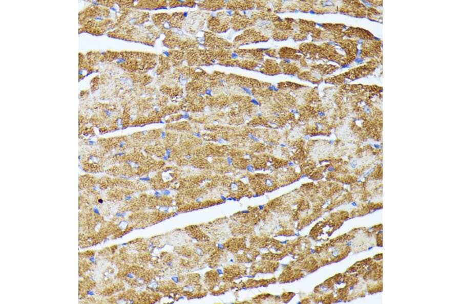
Immunohistochemistry analysis of paraffin-embedded mouse heart using Anti-TRAP1 Antibody (A14167) at a dilution of 1:100 (40x lens). Perform microwave antigen retrieval with 10 mM PBS buffer pH 7.2 before commencing with IHC staining protocol.
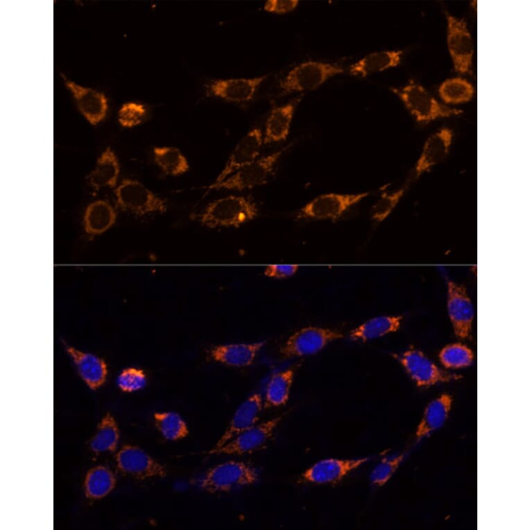
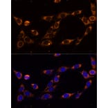
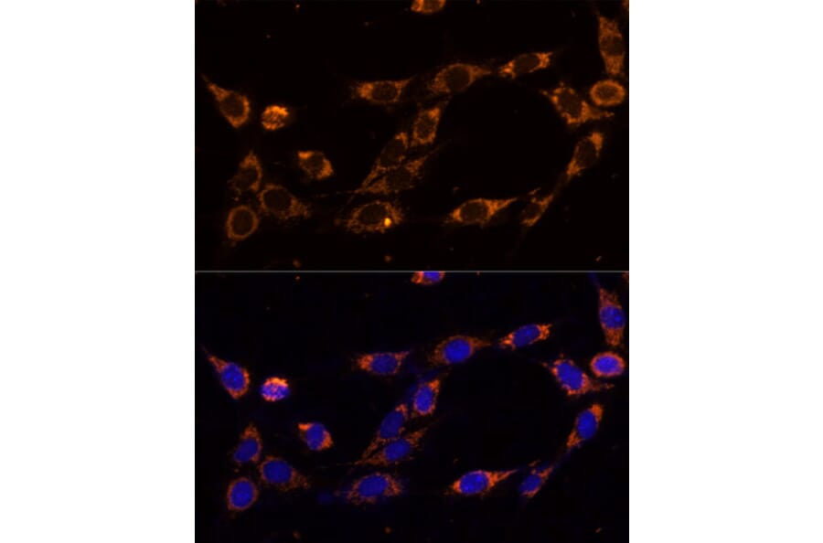
Immunofluorescence analysis of NIH-3T3 cells using Anti-TRAP1 Antibody (A14167) at a dilution of 1:100 (40x lens). DAPI was used to stain the cell nuclei (blue).
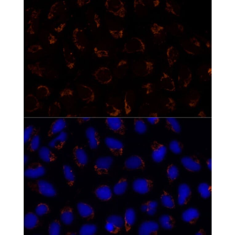
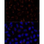
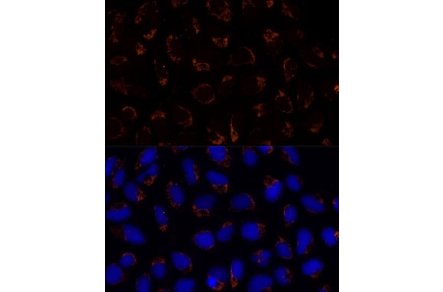
Immunofluorescence analysis of U-2 OS cells using Anti-TRAP1 Antibody (A14167) at a dilution of 1:100 (40x lens). DAPI was used to stain the cell nuclei (blue).