Immunocytochemistry/Immunofluorescence analysis of human HaCaT cells, fixed in cold 100% methanol for 10 minutes at -20°C, using Anti-TRPV3 Antibody [N15/39] (A305015), at 1:100 for 1 hour at room temperature. The secondary antibody used was FITC Goat Anti-Mouse (green) at 1:50 for 1 hour at room temperature. Localization: Dotty staining in all cells. Some intermediate filament-like staining in some cells.
Western Blot - Anti-TRPV3 Antibody [N15/39] (A305015)
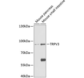
Western blot analysis of rat brain membrane lysate showing detection of TrpV3 protein using Anti-TRPV3 Antibody [N15/39] (A305015) at 1:1,000 for 2 hours at room temperature. Load: 15 µg. Block: 1.5% BSA for 30 minutes at room temperature. The secondary antibody used was Sheep Anti-Mouse IgG: HRP for 1 hour at room temperature.
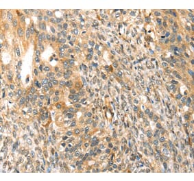
Immunohistochemistry analysis of mouse backskin, fixed in Bouin's fixative solution and paraffin-embedded. The Primary Antibody used was Anti-TRPV3 Antibody [N15/39] (A305015) at 1:100 for 1 hour at room temperature. The secondary antibody used was FITC Goat Anti-Mouse (green) at 1:50 for 1 hour at room temperature. Localization: Filaggrin-like staining in upper layers. Dull lower layer cell staining. Some stain seen in hypodermis.
Immunocytochemistry/Immunofluorescence analysis of human neuroblastoma cells (SH-SY5Y), fixed in 4% PFA for 15 min, using Anti-TRPV3 Antibody [N15/39] (A305015), at 1:50 for overnight at 4°C with slow rocking. The secondary antibody used was AlexaFluor 488 at 1:1,000 for 1 hour at room temperature. Counterstain: Phalloidin-iFluor 647 (red) F-Actin stain; Hoechst (blue) nuclear stain at 1:800, 1.6mM for 20 minutes at room temperature.(A) Hoechst (blue) nuclear stain. (B) Phalloidin-iFluor 647 (red) F-Actin stain. (C) TrpV3 Antibody (D) Composite.
Publishing research using Anti-TRPV3 Antibody [N15/39] (A305015)? Please let us know so that we can list the citation on this page.
Alternative products to Anti-TRPV3 Antibody [N15/39] (A305015)


![Immunocytochemistry/Immunofluorescence - Anti-TRPV3 Antibody [N15/39] (A305015) - Antibodies.com](https://cdn.antibodies.com/image/catalog/305/A305015_1.png?profile=product_top)
![Western Blot - Anti-TRPV3 Antibody [N15/39] (A305015) - Antibodies.com](https://cdn.antibodies.com/image/catalog/305/A305015_2.png?profile=product_top)
![Immunohistochemistry - Anti-TRPV3 Antibody [N15/39] (A305015) - Antibodies.com](https://cdn.antibodies.com/image/catalog/305/A305015_3.png?profile=product_top)
![Immunocytochemistry/Immunofluorescence - Anti-TRPV3 Antibody [N15/39] (A305015) - Antibodies.com](https://cdn.antibodies.com/image/catalog/305/A305015_4.png?profile=product_top)
![Immunocytochemistry/Immunofluorescence - Anti-TRPV3 Antibody [N15/39] (A305015) - Antibodies.com](https://cdn.antibodies.com/image/catalog/305/A305015_1.png?profile=product_top_thumb)
![Western Blot - Anti-TRPV3 Antibody [N15/39] (A305015) - Antibodies.com](https://cdn.antibodies.com/image/catalog/305/A305015_2.png?profile=product_top_thumb)
![Immunohistochemistry - Anti-TRPV3 Antibody [N15/39] (A305015) - Antibodies.com](https://cdn.antibodies.com/image/catalog/305/A305015_3.png?profile=product_top_thumb)
![Immunocytochemistry/Immunofluorescence - Anti-TRPV3 Antibody [N15/39] (A305015) - Antibodies.com](https://cdn.antibodies.com/image/catalog/305/A305015_4.png?profile=product_top_thumb)
![Immunocytochemistry/Immunofluorescence - Anti-TRPV3 Antibody [N15/39] (A305015) - Antibodies.com](https://cdn.antibodies.com/image/catalog/305/A305015_1.png?profile=product_image)
![Western Blot - Anti-TRPV3 Antibody [N15/39] (A305015) - Antibodies.com](https://cdn.antibodies.com/image/catalog/305/A305015_2.png?profile=product_image)
![Immunohistochemistry - Anti-TRPV3 Antibody [N15/39] (A305015) - Antibodies.com](https://cdn.antibodies.com/image/catalog/305/A305015_3.png?profile=product_image)
![Immunocytochemistry/Immunofluorescence - Anti-TRPV3 Antibody [N15/39] (A305015) - Antibodies.com](https://cdn.antibodies.com/image/catalog/305/A305015_4.png?profile=product_image)
![Immunocytochemistry/Immunofluorescence - Anti-TRPV3 Antibody [N15/4] (A305016) - Antibodies.com](https://cdn.antibodies.com/image/catalog/305/A305016_1.png?profile=product_alternative)