Supplied in Phosphate Buffered Saline, pH 7.3, with 50% Glycerol and 0.05% Proclin 300.
Storage
Shipped at 4°C. Upon delivery aliquot and store at -20°C. Avoid freeze/thaw cycles.
Synonyms
E3 UFM1-protein ligase 1, E3 UFM1-protein transferase 1, KIAA0776, MAXER, Multiple alpha-helix protein located at ER, NLBP, Novel LZAP-binding protein, RCAD, Regulator of C53/LZAP and DDRGK1
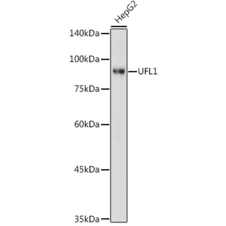
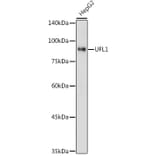
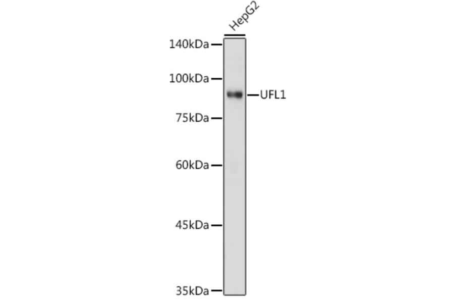
Western blot analysis of extracts of HepG2 cells, using Anti-UFL1 Antibody (A305259) at 1:1,000 dilution. The secondary antibody was Goat Anti-Rabbit IgG H&L Antibody (HRP) at 1:10,000 dilution. Lysates/proteins were present at 25µg per lane. The blocking buffer used was 3% non-fat dry milk in TBST. Detection was with a ECL Basic Kit. Exposure time: 1s.
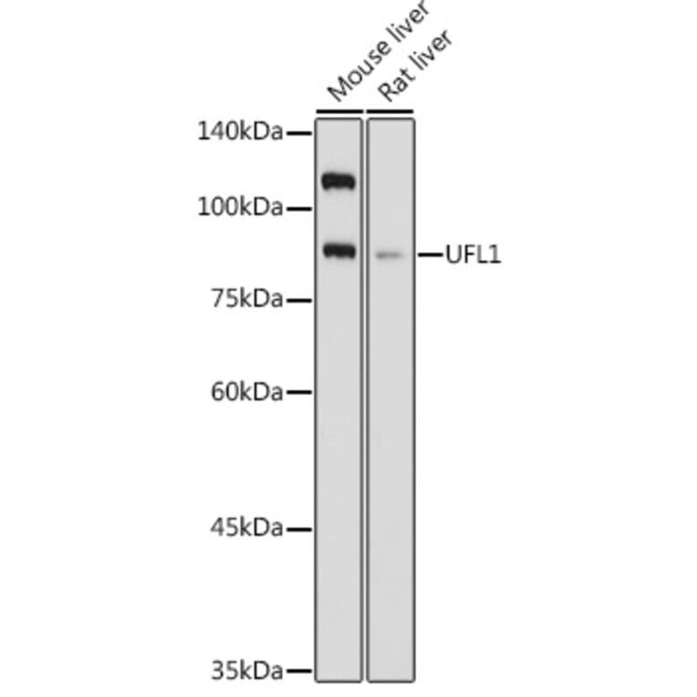
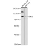
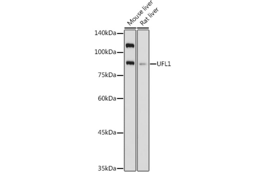
Western blot analysis of extracts of various cell lines, using Anti-UFL1 Antibody (A305259) at 1:1,000 dilution. The secondary antibody was Goat Anti-Rabbit IgG H&L Antibody (HRP) at 1:10,000 dilution. Lysates/proteins were present at 25µg per lane. The blocking buffer used was 3% non-fat dry milk in TBST. Detection was with a ECL Basic Kit. Exposure time: 3s.
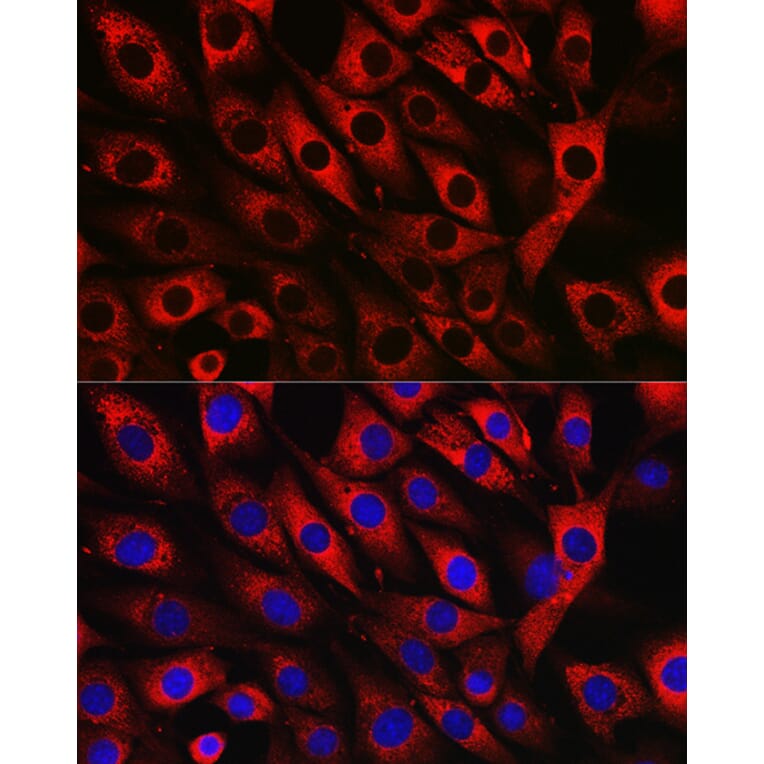
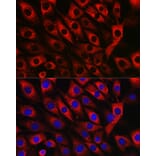
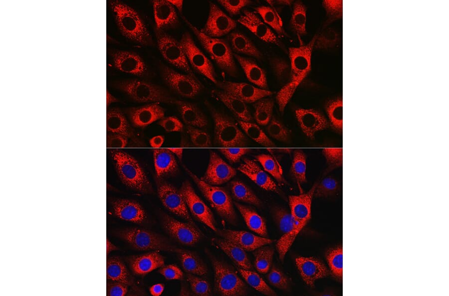
Immunofluorescence analysis of NIH/3T3 cells using Anti-UFL1 Antibody (A305259) at a dilution of 1:50 (40x lens). DAPI was used to stain the cell nuclei (blue).
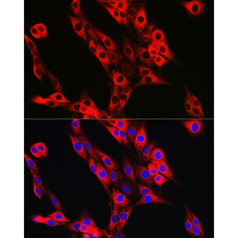
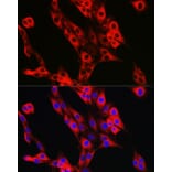
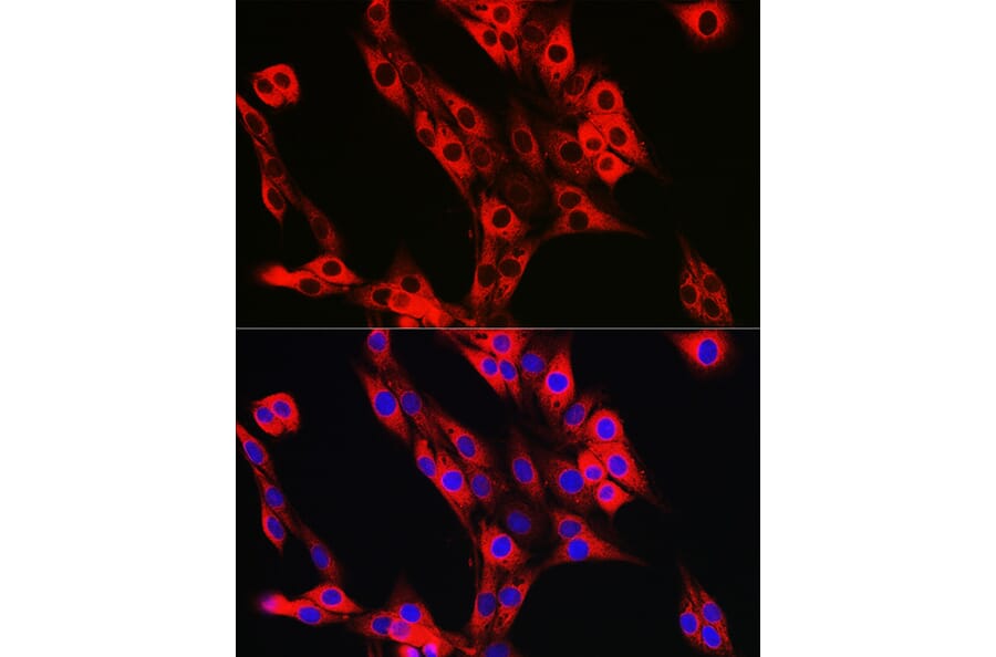
Immunofluorescence analysis of PC-12 cells using Anti-UFL1 Antibody (A305259) at a dilution of 1:50 (40x lens). DAPI was used to stain the cell nuclei (blue).
Publishing research using Anti-UFL1 Antibody (A305259)? Please let us know so that we can list the citation on this page.
Proteins predicted to interact with UFL1
Predicted protein interactions based upon String database. Revelancy score correlates with probability of interaction.