This antibody targets the C-terminal domain of human VIP Receptor 1, recognizing the non-phosphorylated form. It is validated for Western blot analysis, supports immunoprecipitation from cell and tissue lysates, and is suitable for immunohistochemistry in cultured cells and tissue sections.
Applications
WB, ICC, IHC
Dilutions
WB: 1,000, IHC: 1:100, ICC: 1:200
Reactivity
Human
Immunogen
Synthetic peptide corresponding to human VIP Receptor 1 (amino acids. 438-457). Immunogen range is 22-22 amino acids.
Sequence
TRVSPGARRSSSFQAEVSLV
Host
Rabbit
Clonality
Polyclonal
Isotype
IgG
Conjugate
Unconjugated
Purification
Antigen affinity purification.
Concentration
Lot Specific
Product Form
Liquid
Formulation
Supplied in Dulbecco's PBS, pH 7.4, with 150 mM NaCl and 0.005% Sodium Azide.
Storage
Shipped at 4°C. Upon delivery aliquot and store at -20°C. Avoid freeze/thaw cycles.
Synonyms
PACAP type II receptor, PACAP-R-2, PACAP-R2, Pituitary adenylate cyclase-activating polypeptide type II receptor, Vasoactive intestinal polypeptide receptor 1, VIP-R-1, VIPR1, VPAC1
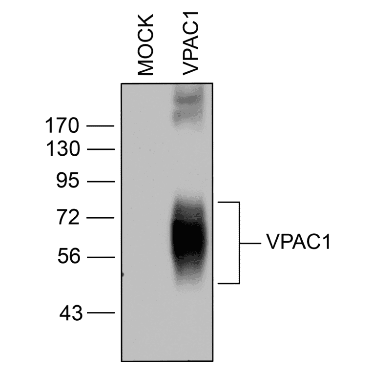
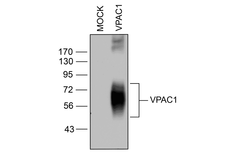
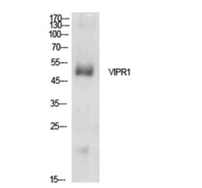
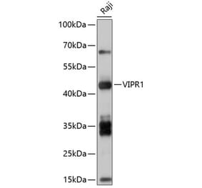
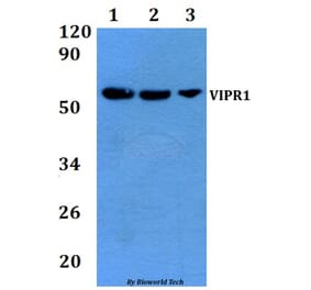
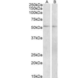
Western blot confirmation of VIP Receptor 1 (VIP Receptor 1) in transfected HEK293 cells. Lysates from native HEK293 cells (MOCK) or cells stably expressing VIP Receptor 1 were probed with anti-VIP Receptor 1 antibody (A334554) at 1:1000.
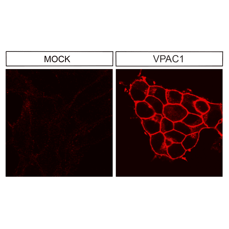
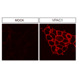
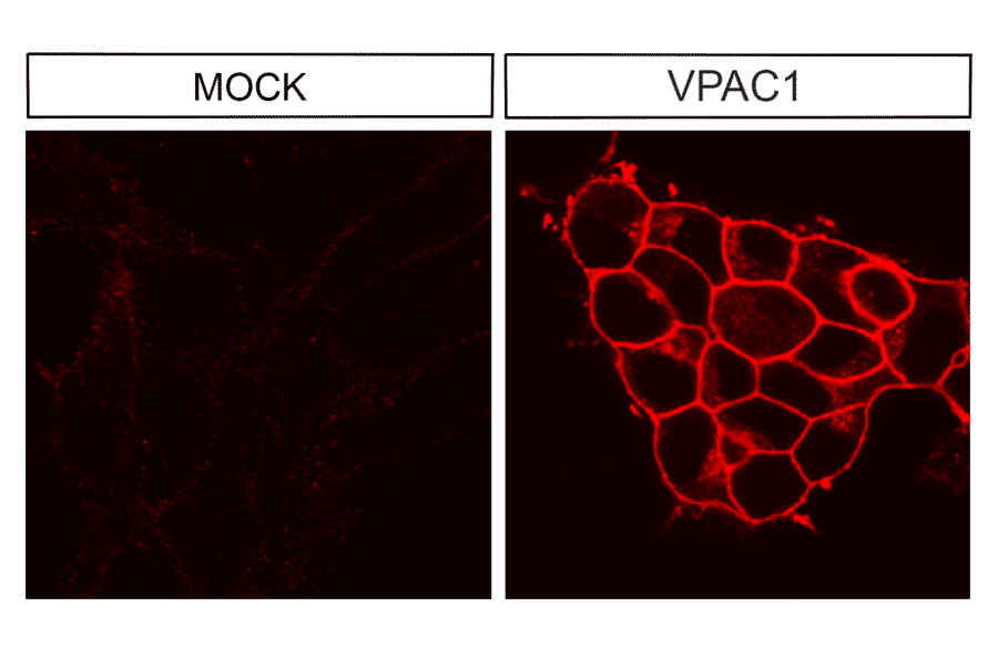
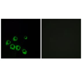
Immunocytochemical analysis of VIP Receptor 1 (VIP Receptor 1) in transfected HEK293 cells. Native HEK293 cells (MOCK) or cells stably expressing VIP Receptor 1 were stained with anti-VIP Receptor 1 antibody (A334554) at 1:200. VIP Receptor 1 receptors were observed at the plasma membrane in transfected cells.
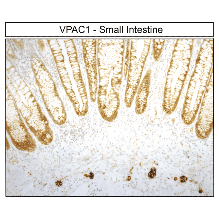
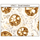
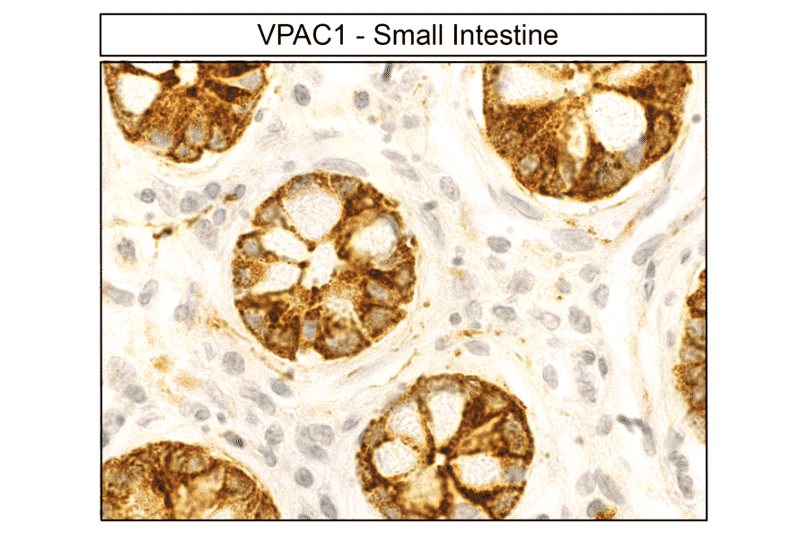
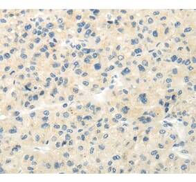
Immunohistochemical detection of VIP Receptor 1 (VIP Receptor 1) in human small intestine. Small intestine sections were dewaxed, microwaved in citric acid, and stained with anti-VIP Receptor 1 antibody (A334554) at 1:100, followed by biotinylated anti-rabbit IgG and avidin-biotin solution. Sections were developed with 3,3-diaminobenzidine (DAB)-glucose oxidase and lightly counterstained with hematoxylin. VIP Receptor 1 receptors were uniformly detected at the plasma membrane of epithelial cells.
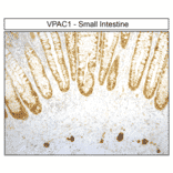
Localization of VIP Receptor 1 (VIP Receptor 1) in human small intestine by immunohistochemistry. Small intestine sections were dewaxed, microwaved in citric acid, and incubated with anti-VIP Receptor 1 antibody (A334554) at 1:100, followed by biotinylated anti-rabbit IgG and avidin-biotin solution. Sections were developed with 3,3-diaminobenzidine (DAB)-glucose oxidase and lightly counterstained with hematoxylin. VIP Receptor 1 receptor staining was absent in the mucosa but present in the basal portion of the crypts.
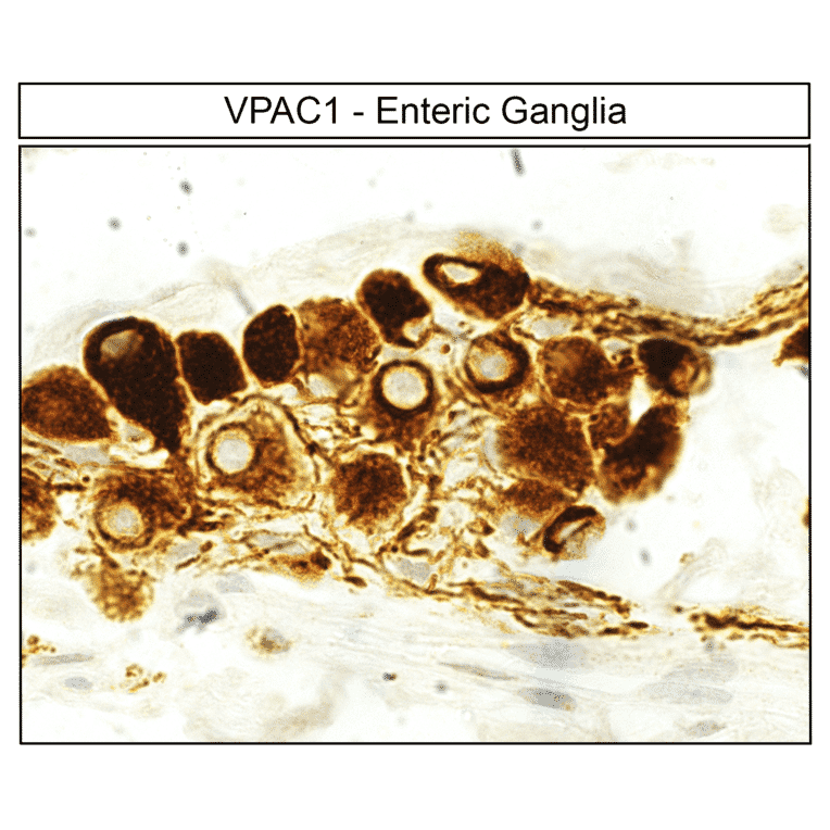
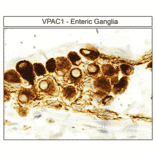
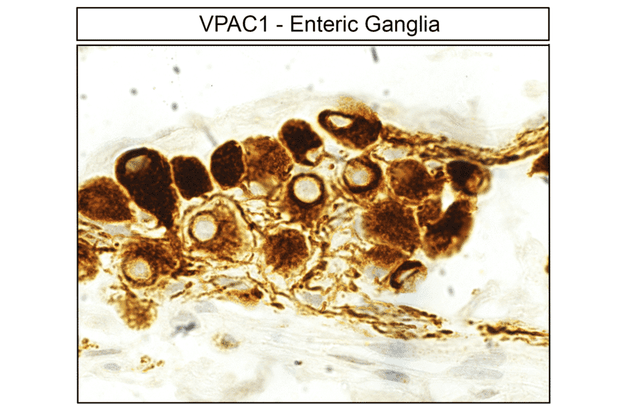
Immunohistochemical detection of VIP Receptor 1 (VIP Receptor 1) in human enteric ganglia. Enteric ganglia sections were dewaxed, microwaved in citric acid, and stained with anti-VIP Receptor 1 antibody (A334554) at 1:100, followed by biotinylated anti-rabbit IgG and avidin-biotin solution. Sections were developed with 3,3-diaminobenzidine (DAB)-glucose oxidase and lightly counterstained with hematoxylin. VIP Receptor 1 receptors were uniformly detected in nearly all enteric ganglia cells.