This antibody specifically recognizes the C-terminal region of human VPAC2, detecting the non-phosphorylated form. It is optimized for Western blot analysis, supports immunoprecipitation from cell and tissue lysates, and is suitable for immunohistochemistry in cultured cells and tissue sections.
Applications
WB, ICC, IHC
Dilutions
WB: 1,000, IHC: 1:100, ICC: 1:200
Reactivity
Human, Mouse, Rat
Immunogen
Synthetic peptide corresponding to VPAC2 (amino acids. 419-438). Immunogen range is 22-22 amino acids.
Sequence
LQFHRGSRAQSFLQTETSVI
Host
Rabbit
Clonality
Polyclonal
Isotype
IgG
Conjugate
Unconjugated
Purification
Antigen affinity purification.
Concentration
Lot Specific
Product Form
Liquid
Formulation
Supplied in Dulbecco's PBS, pH 7.4, with 150 mM NaCl and 0.005% Sodium Azide.
Storage
Shipped at 4°C. Upon delivery aliquot and store at -20°C. Avoid freeze/thaw cycles.
Synonyms
Helodermin-preferring VIP receptor, PACAP type III receptor, PACAP-R-3, PACAP-R3, Pituitary adenylate cyclase-activating polypeptide type III receptor, Vasoactive intestinal polypeptide receptor 2, VIP-R-2, VIP2R, VIPR2
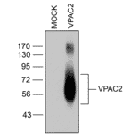
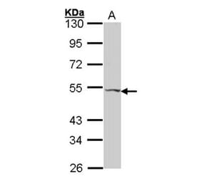
Western blot validation of VIP Receptor 2 (VPAC2) in transfected HEK293 cells. Lysates from native HEK293 cells (MOCK) or cells stably expressing VPAC2 were probed with phosphorylation-independent anti-VPAC2 antibody (A334555) at 1:1000.
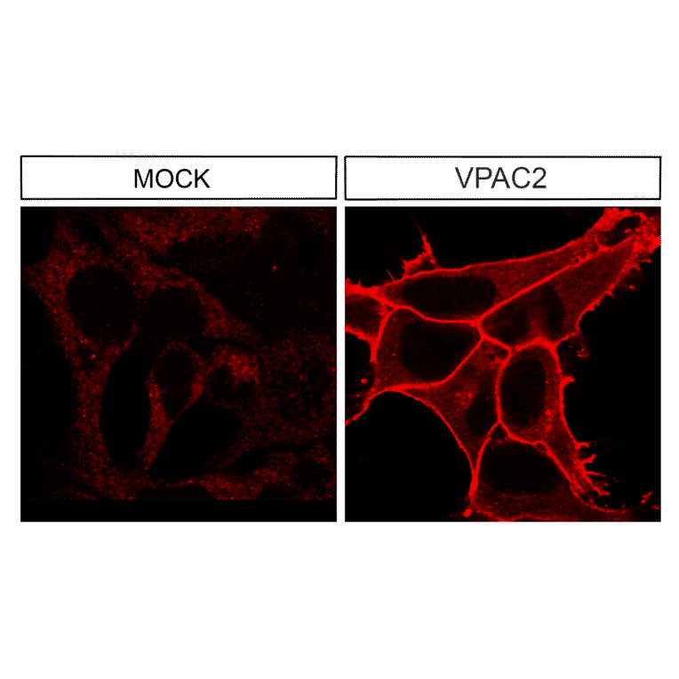
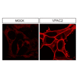
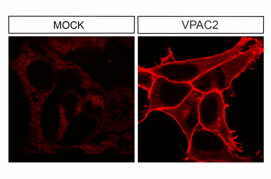
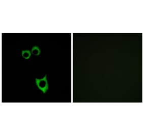
Immunocytochemical analysis of VIP Receptor 2 (VPAC2) in transfected HEK293 cells. Native HEK293 cells (MOCK) or cells stably expressing VPAC2 were stained with anti-VPAC2 antibody (A334555) at 1:200. VPAC2 receptors were confined to the plasma membrane in transfected cells.
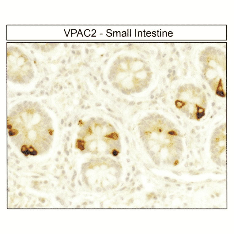
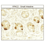
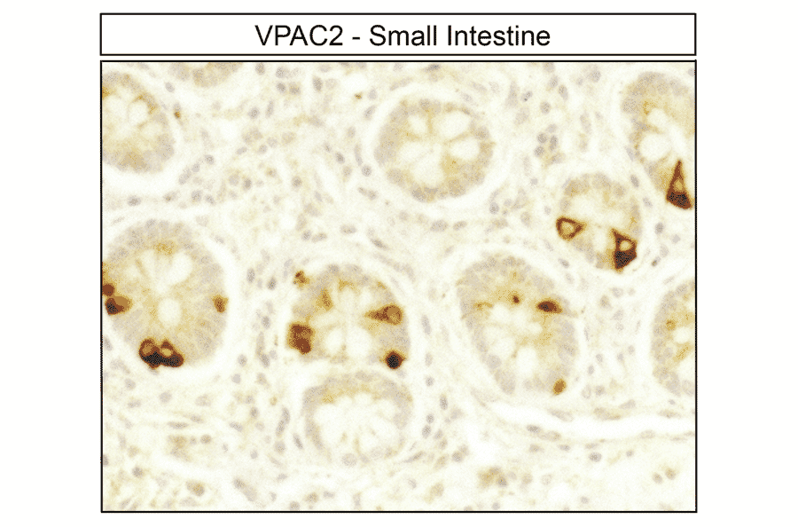
Immunohistochemical localization of VIP Receptor 2 (VPAC2) in human small intestine. Small intestine sections were dewaxed, microwaved in citric acid, and stained with anti-VPAC2 antibody (A334555) at 1:100, followed by biotinylated anti-rabbit IgG and avidin-biotin solution. Color was developed with 3-amino-9-ethylcarbazole (AEC), and sections were counterstained with hematoxylin. VPAC2 receptors were detected at the plasma membrane of neuroendocrine cells.
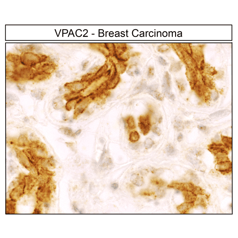
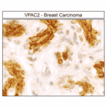
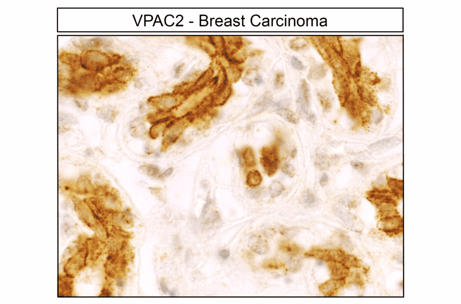
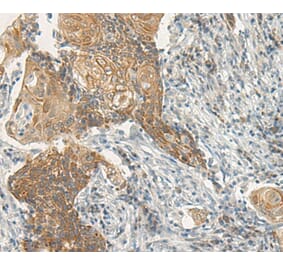
Detection of VIP Receptor 2 (VPAC2) in human breast carcinoma by immunohistochemistry. Breast carcinoma sections were dewaxed, microwaved in citric acid, and incubated with anti-VPAC2 antibody (A334555) at 1:100, followed by biotinylated anti-rabbit IgG and avidin-biotin solution. Sections were developed with 3,3-diaminobenzidine (DAB)-glucose oxidase and lightly counterstained with hematoxylin. VPAC2 receptors were uniformly detected at the plasma membrane of nearly all tumor cells.
Alternative products to Anti-VPAC2 Antibody (A334555)