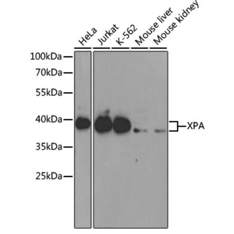
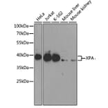
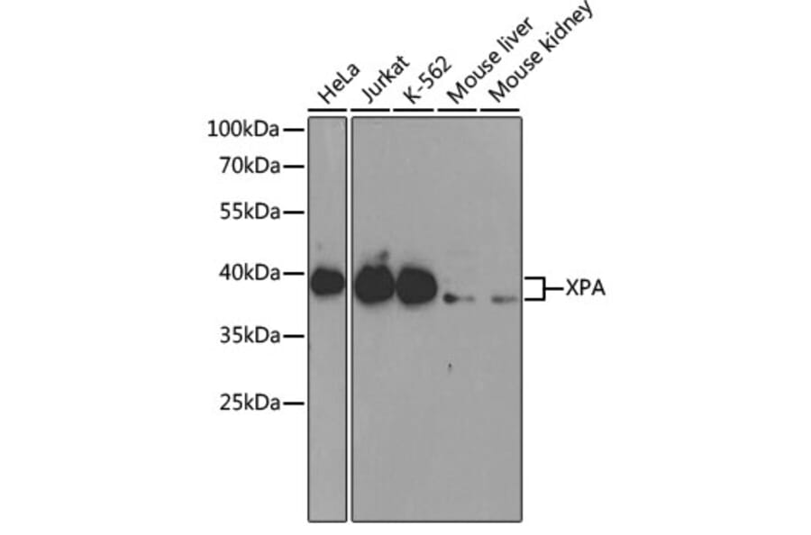
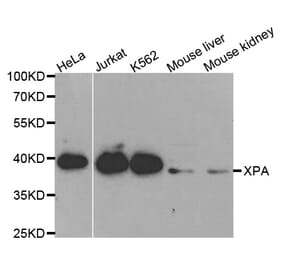
Western blot analysis of extracts of various cell lines, using Anti-XPA Antibody (A1626) at 1:1,000 dilution. Secondary antibody: Goat Anti-Rabbit IgG (H+L) (HRP) (AS014) at 1:10,000 dilution. Lysates / proteins: 25µg per lane. Blocking buffer: 3% non-fat dry milk in TBST.
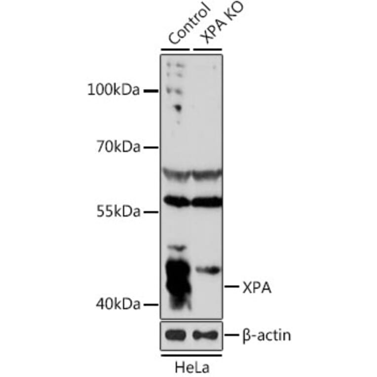
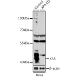
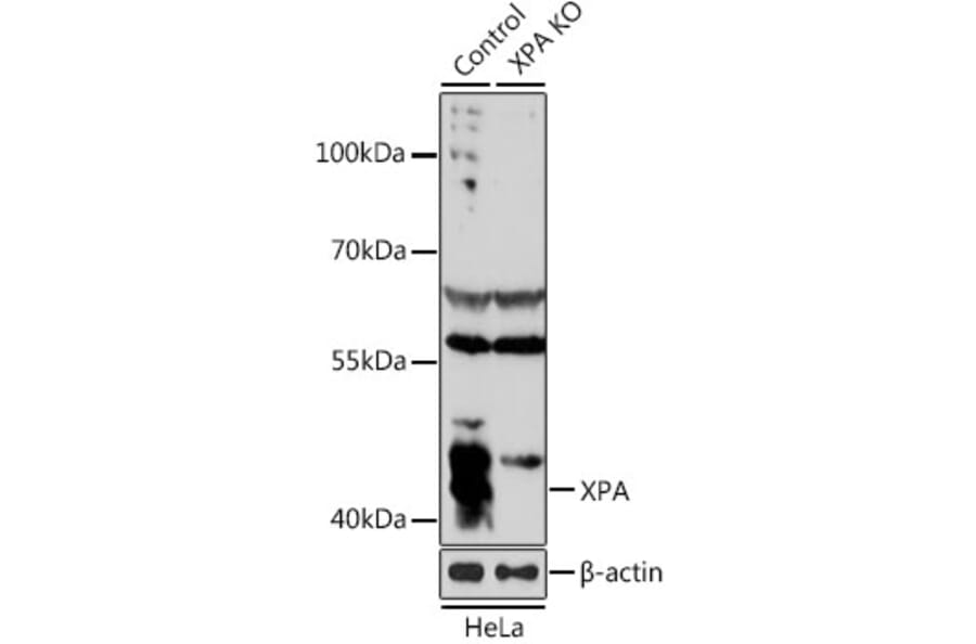
Western blot analysis of extracts from normal (control) and XPA knockout (KO) HeLa cells, using Anti-XPA Antibody (A1626) at 1:1,000 dilution. Secondary antibody: Goat Anti-Rabbit IgG (H+L) (HRP) (AS014) at 1:10,000 dilution. Lysates / proteins: 25µg per lane. Blocking buffer: 3% non-fat dry milk in TBST. Detection: ECL Basic Kit (RM00020). Exposure time: 15s.
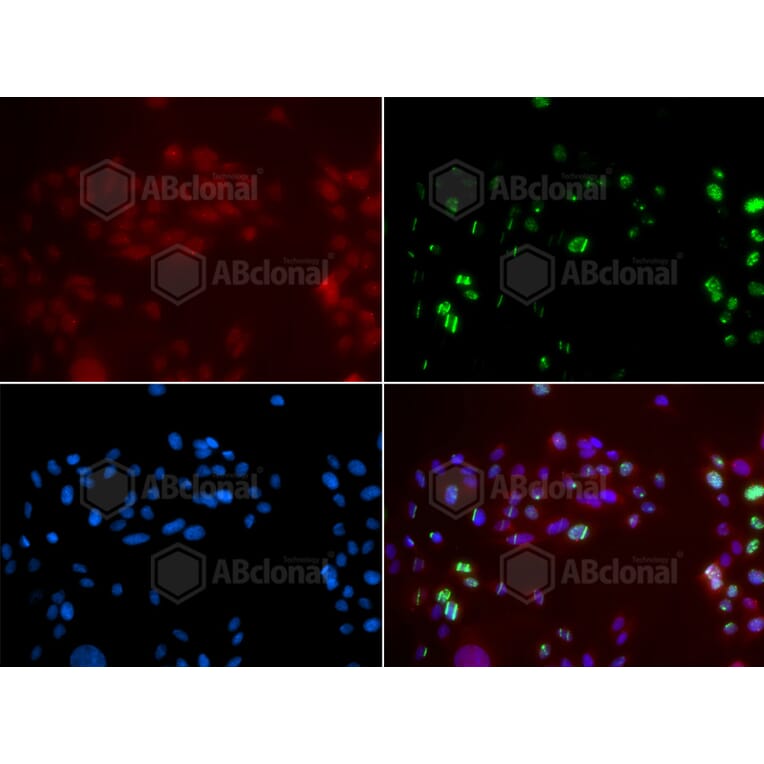
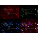
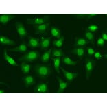
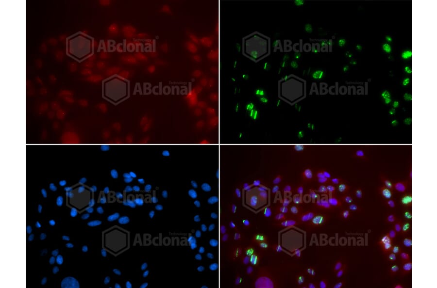
Immunofluorescence analysis of GFP-RNF168 transgenic U2OS cells using Anti-XPA Antibody (A1626). Green: GFP-RNF168 fusion protein expression for DNA damage marker.Blue: DAPI for nuclear staining.RNF168 (GFP) can be used to mark cells damaged by UV-A laser for they always gather around DNA damage region.