This product recognizes the heavy and light chains of Mouse IgG.
Applications
ICC/IF, Flow Cytometry
Recommended Dilutions
IF: 2 drops/mL, Flow Cytometry: 1 drop/mL
Clonality
Polyclonal
Isotype
IgG
Conjugate
CF®543
Product Form
Liquid
Formulation
Supplied in Phosphate Buffered Saline with 50% Glycerol, 2 mg/mL BSA and 0.05% Sodium Azide.
Storage
Shipped and stored at +4°C. This product is also photosensitive and should be protected from light. CF® Dyes are guaranteed for at least 6 months from data of receipt when stored correctly.
General Notes
Looking for a specific protein conjugate to simplify your workflow? We offer a library of over 2,000 targets conjugated to your choice of CF® dye. To enquire about a custom product, contact us directly.
Disclaimer
This product is for research use only. It is not intended for diagnostic or therapeutic use.
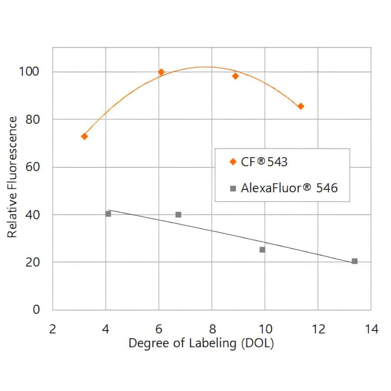
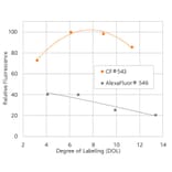
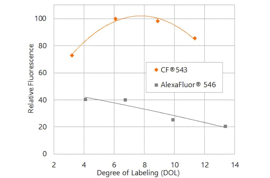
CF®543 produces substantially brighter antibody conjugates than Alexa Fluor® 546. Relative fluorescence intensity of goat anti-mouse secondary antibody conjugates is shown as a function of degree of labeling (DOL, dye molecules per antibody).
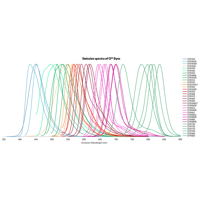
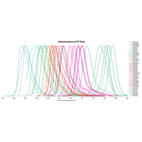
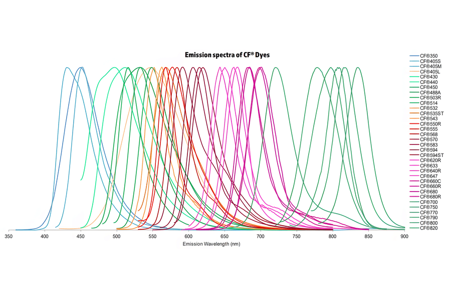
Normalized emission spectra of the CF® dye family spanning the visible to near-infrared range are shown, illustrating the spectral diversity and overlap between dyes. Curves represent relative fluorescence intensity as a function of emission wavelength (nm), with peak positions corresponding to each dye’s characteristic emission maximum. This reference highlights the broad coverage of CF® dyes for multicolor fluorescence applications and aids in selecting compatible dye combinations for imaging, flow cytometry, and other fluorescence-based assays.
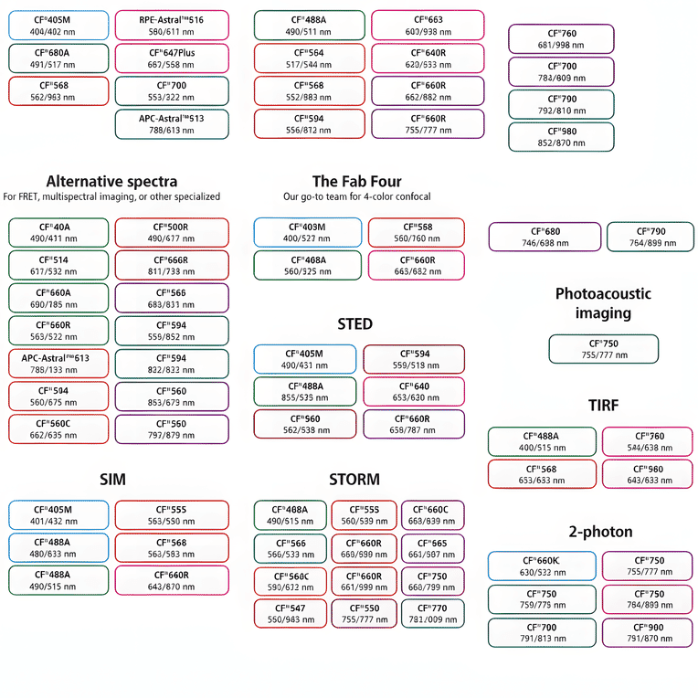
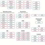
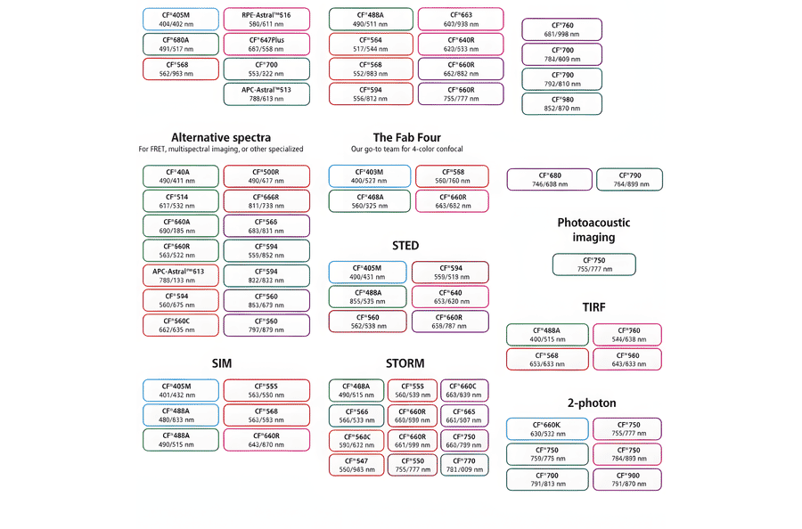
This chart summarizes commonly used CF® dyes grouped by their suitability for specific imaging modalities, including alternative spectra applications, four-color confocal imaging, near-infrared western blotting, photoacoustic imaging, STED, SIM, STORM, TIRF, and two-photon microscopy. Each dye is shown with its characteristic excitation and emission wavelengths (nm), providing a practical reference for selecting spectrally compatible dyes and optimizing multicolor experimental design across a range of fluorescence techniques.
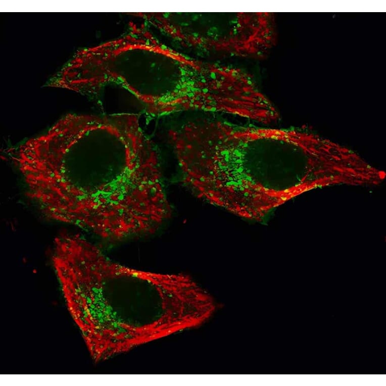
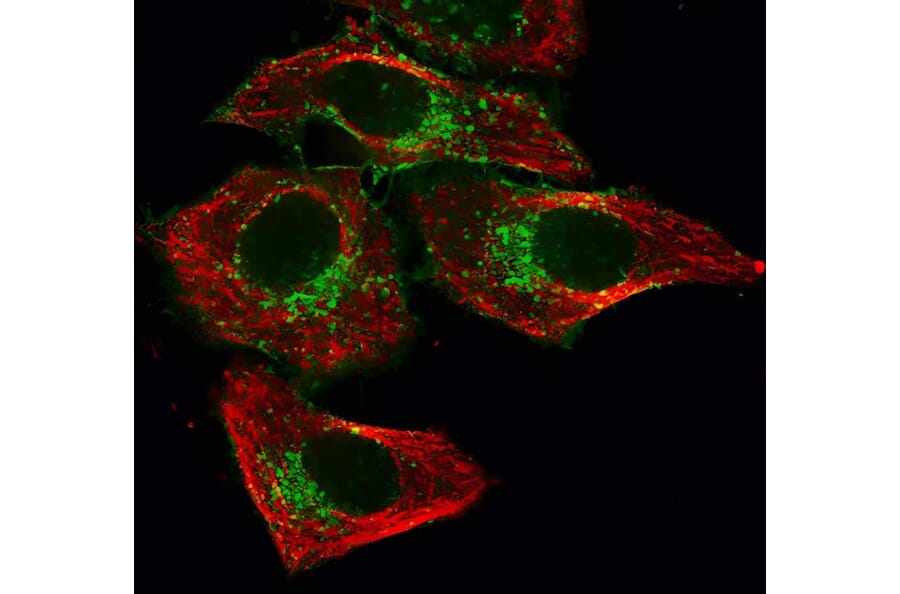
HeLa cells stained with mouse anti-tubulin followed by CF®543 goat anti-mouse to label microtubules (red), and CF®488A Mix-n-Stain™–labeled mouse anti-transferrin receptor to label endosomes (green).
Publishing research using Donkey Anti-Mouse IgG H&L Antibody (CF®543), Cross-Adsorbed (A343771)? Please let us know so that we can list the citation on this page.