Goat Anti-Mouse IgG Fc Antibody (CF®750), Cross-Adsorbed
Description
Goat Anti-Mouse IgG Fc (CF®750) secondary antibody.
Host Species
Goat
Target Species
Mouse
Specificity
This goat anti-mouse IgG3 secondary antibody was raised against mouse IgG3 and specifically recognizes the Fc region of the mouse IgG3 heavy chain.
Applications
ICC/IF, IHC, WB, Flow Cytometry
Recommended Dilutions
IF: 1-10 µg/mL, WB: 50-100 ng/mL, IHC: 1-10 µg/mL
Clonality
Polyclonal
Isotype
IgG
Conjugate
CF®750
Product Form
Supplied as a liquid; 1 mg vials are lyophilized.
Concentration
2 mg/mL
Purification
The antibody is affinity-purified and cross-adsorbed.
Formulation
Liquid: Supplied in phosphate buffered saline (PBS) containing 50% glycerol, 2 mg/ml BSA, and 0.05% sodium azide. Lyophilized: Supplied in phosphate Buffered Saline with 15 mg/ml BSA and 20 mg/ml trehalose.
Storage
Shipped at +4°C. Upon delivery aliquot and store at -20°C. Avoid freeze/thaw cycles. This product is also photosensitive and should be protected from light. Should this product contain a precipitate we recommend microcentrifugation before use. CF® Dyes are guaranteed for at least 6 months from data of receipt when stored correctly.
General Notes
Looking for a specific protein conjugate to simplify your workflow? We offer a library of over 2,000 targets conjugated to your choice of CF® dye. To enquire about a custom product, contact us directly.
Disclaimer
This product is for research use only. It is not intended for diagnostic or therapeutic use.
Goat Anti-Mouse IgG Fc Antibody (CF®750), Cross-Adsorbed (A343903)
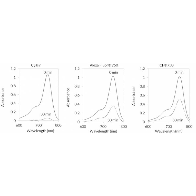
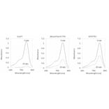
CF®750 demonstrates greater photostability than spectrally similar near-infrared dyes. Shown are the absorbance spectra of NIR fluorophores before light exposure (0 min, solid lines) and after 30 minutes of sunlight exposure (30 min, dashed lines). Following illumination, CF®750 retains higher absorbance compared to Cy®7 and Alexa Fluor® 750, indicating reduced photobleaching and superior resistance to photodegradation.
Goat Anti-Mouse IgG Fc Antibody (CF®750), Cross-Adsorbed (A343903)
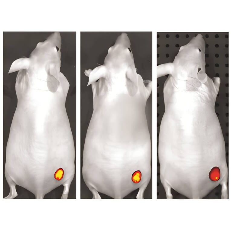
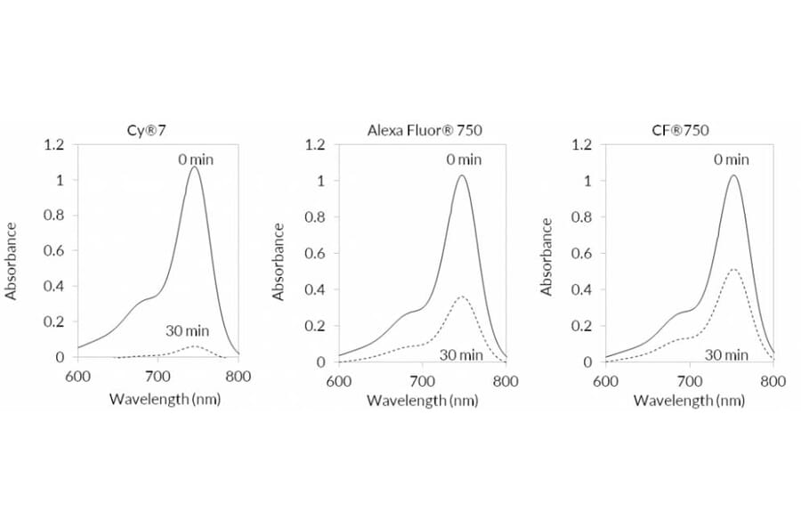
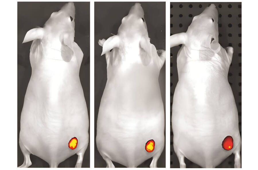
CF®750 for in vivo small animal imaging. Tumor-bearing mice were imaged using an IVIS® imaging system (PerkinElmer) at 24 hours (left), 48 hours (center), and 96 hours (right) following intravenous administration of a CF®750–Avastin® conjugate. Image courtesy of Caliper Life Sciences.
Goat Anti-Mouse IgG Fc Antibody (CF®750), Cross-Adsorbed (A343903)
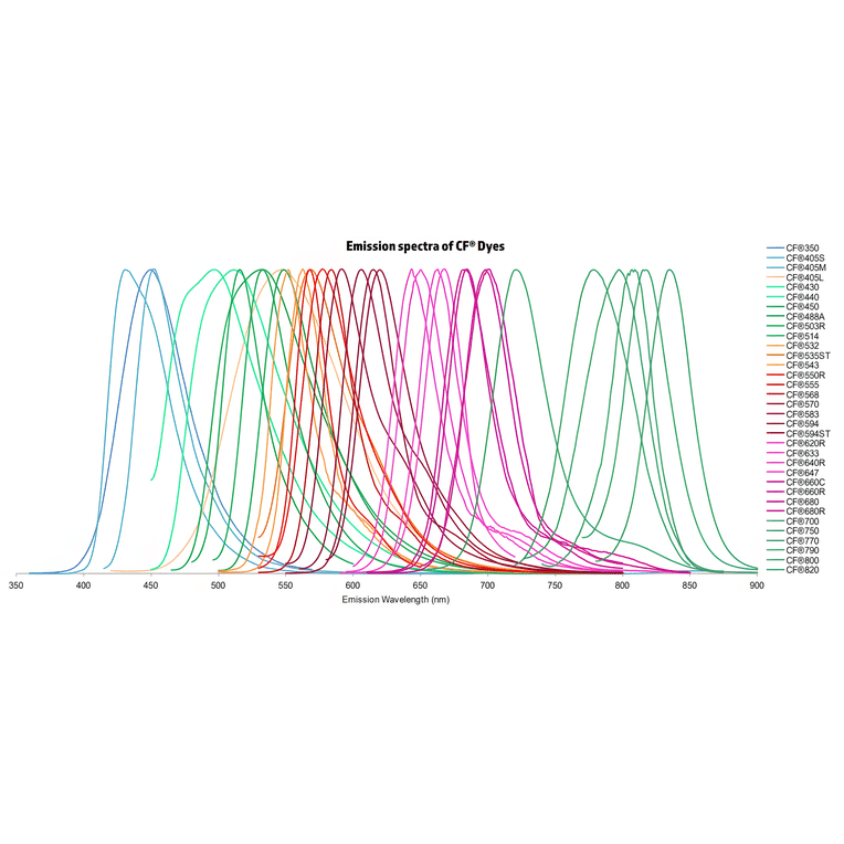
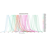
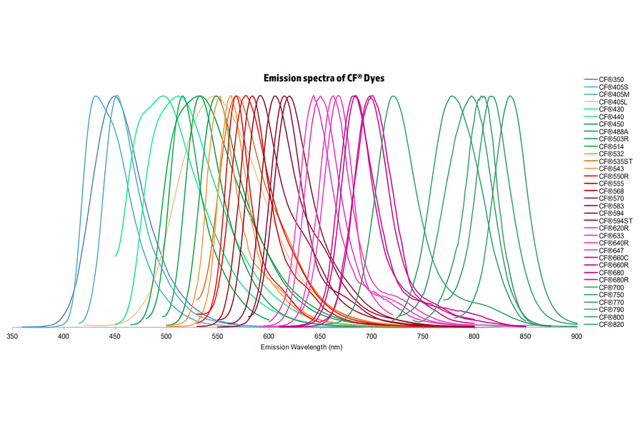
Normalized emission spectra of the CF® dye family spanning the visible to near-infrared range are shown, illustrating the spectral diversity and overlap between dyes. Curves represent relative fluorescence intensity as a function of emission wavelength (nm), with peak positions corresponding to each dye’s characteristic emission maximum. This reference highlights the broad coverage of CF® dyes for multicolor fluorescence applications and aids in selecting compatible dye combinations for imaging, flow cytometry, and other fluorescence-based assays.
Goat Anti-Mouse IgG Fc Antibody (CF®750), Cross-Adsorbed (A343903)
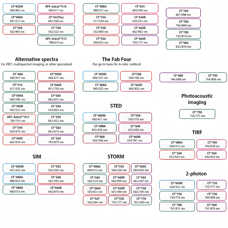
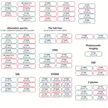
This chart summarizes commonly used CF® dyes grouped by their suitability for specific imaging modalities, including alternative spectra applications, four-color confocal imaging, near-infrared western blotting, photoacoustic imaging, STED, SIM, STORM, TIRF, and two-photon microscopy. Each dye is shown with its characteristic excitation and emission wavelengths (nm), providing a practical reference for selecting spectrally compatible dyes and optimizing multicolor experimental design across a range of fluorescence techniques.
Publishing research using Goat Anti-Mouse IgG Fc Antibody (CF®750), Cross-Adsorbed (A343903)? Please let us know so that we can list the citation on this page.