This product recognizes the heavy and light chains of Goat IgG.
Applications
ICC/IF, IHC, WB, Flow Cytometry
Recommended Dilutions
IF: 1-10 µg/mL, WB: 50-100 ng/mL, IHC: 1-10 µg/mL
Clonality
Polyclonal
Isotype
IgG
Conjugate
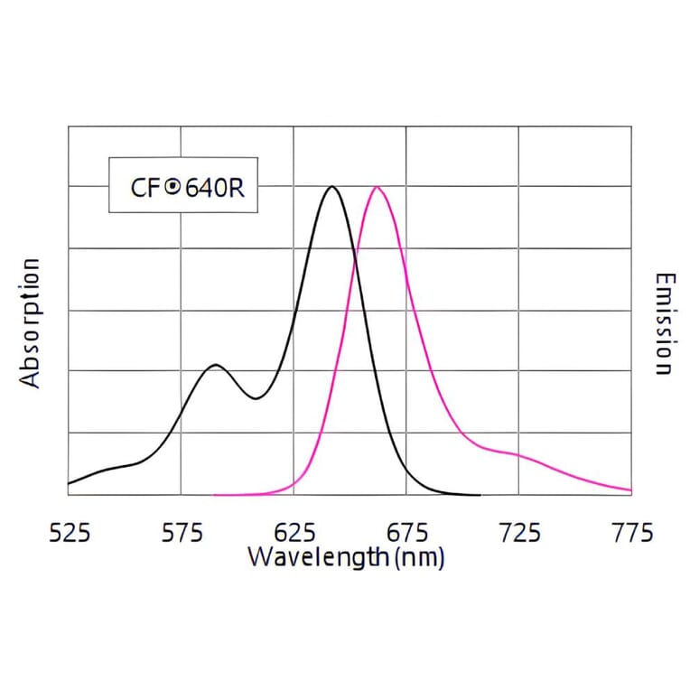
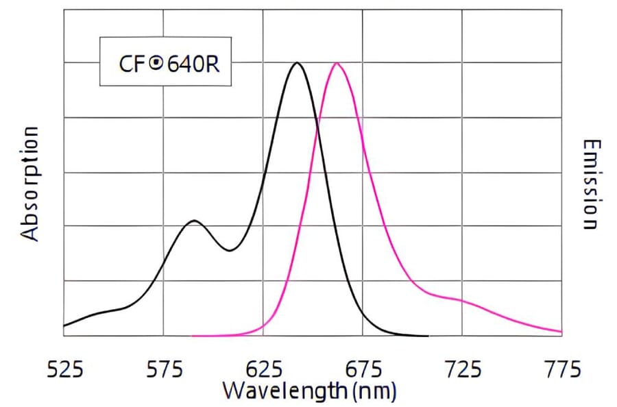
CF®640R
Product Form
Supplied as a liquid; 1 mg vials are lyophilized.
Concentration
2 mg/mL
Formulation
Liquid: Supplied in Phosphate Buffered Saline containing 50% glycerol, 2 mg/ml BSA, and 0.05% Sodium Azide
Storage
Shipped at +4°C. Upon delivery aliquot and store at -20°C. Avoid freeze/thaw cycles. This product is also photosensitive and should be protected from light. Should this product contain a precipitate we recommend microcentrifugation before use. CF® Dyes are guaranteed for at least 6 months from data of receipt when stored correctly.
General Notes
Looking for a specific protein conjugate to simplify your workflow? We offer a library of over 2,000 targets conjugated to your choice of CF® dye. To enquire about a custom product, contact us directly.
Disclaimer
This product is for research use only. It is not intended for diagnostic or therapeutic use.
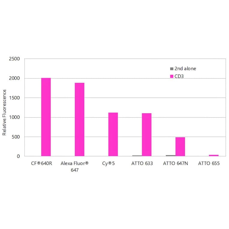
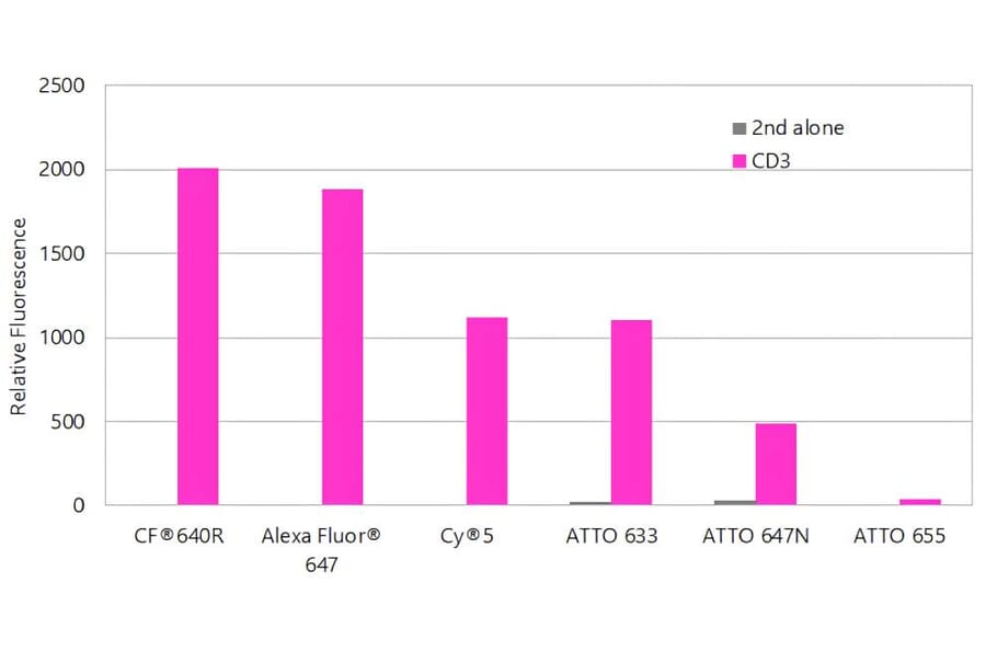
CF®640R produces brighter antibody conjugates than other far-red dyes. Jurkat cells were stained with mouse anti-CD3 primary antibody or without primary antibody as a control, followed by goat anti-mouse secondary antibodies conjugated to the indicated dyes. Fluorescence was analyzed on a BD FACSCalibur™ flow cytometer using the FL4 detection channel, and bars represent geometric mean fluorescence intensity.
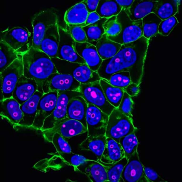
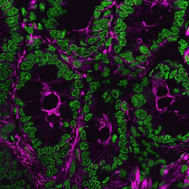
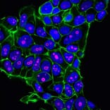
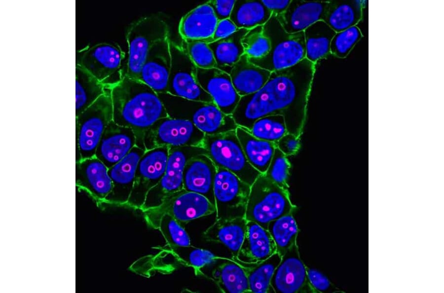
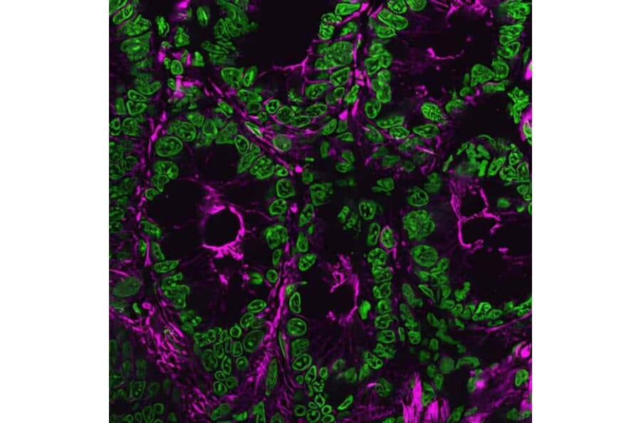
MCF-7 cells stained with CF®640R-conjugated anti-Cyclin B1 antibody to label nuclei and nucleoli (magenta), CF®488A phalloidin to visualize actin filaments (green), and Hoechst to stain DNA (blue).
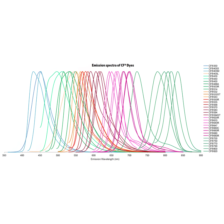
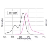
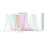
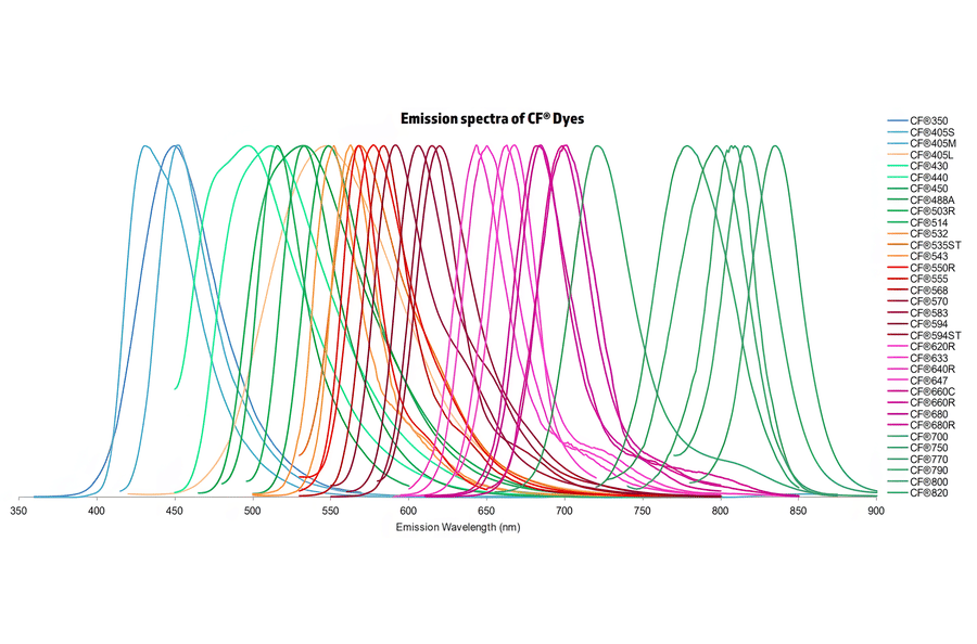
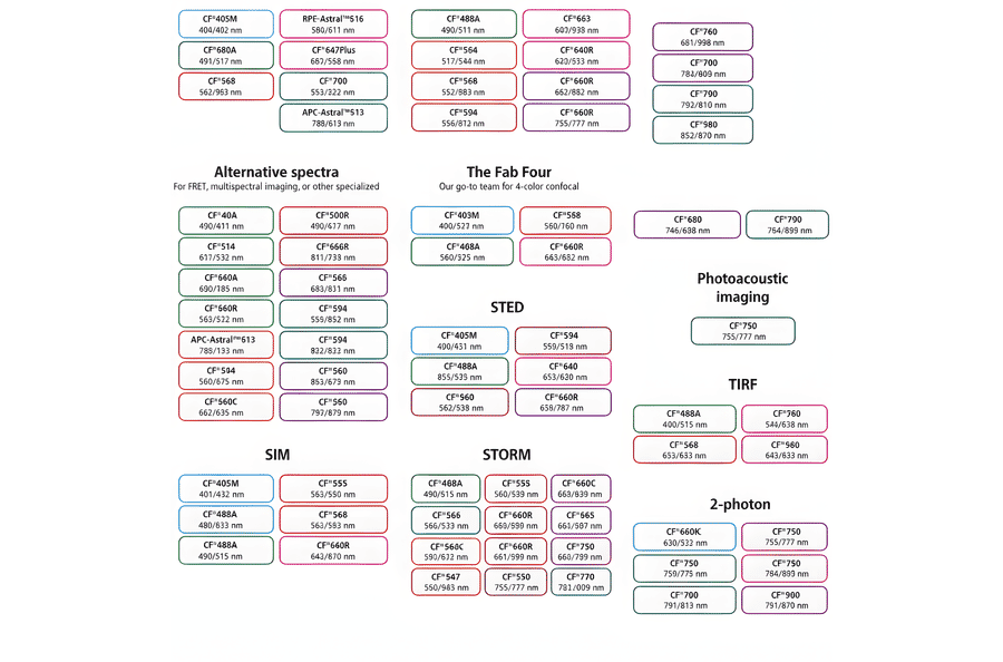
Normalized emission spectra of the CF® dye family spanning the visible to near-infrared range are shown, illustrating the spectral diversity and overlap between dyes. Curves represent relative fluorescence intensity as a function of emission wavelength (nm), with peak positions corresponding to each dye’s characteristic emission maximum. This reference highlights the broad coverage of CF® dyes for multicolor fluorescence applications and aids in selecting compatible dye combinations for imaging, flow cytometry, and other fluorescence-based assays.
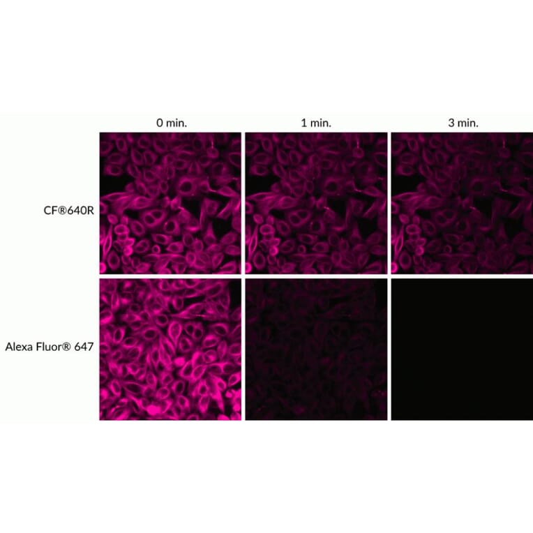
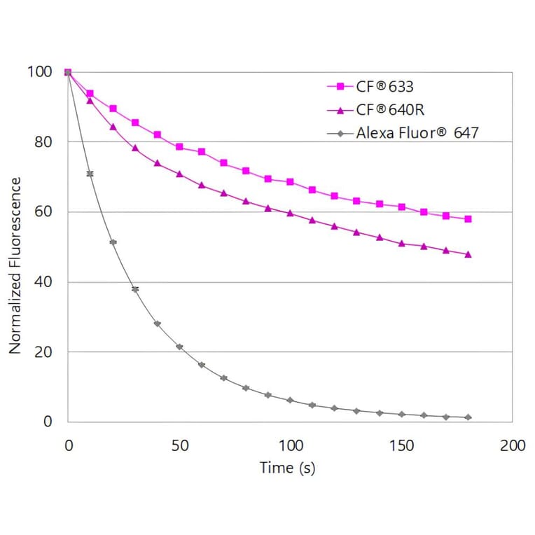
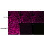
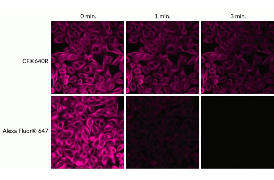
Relative photostability of CF®640R and Alexa Fluor® 647 fluorescence. HeLa cells were stained with mouse anti-tubulin primary antibody followed by goat anti-mouse secondary antibodies conjugated to the indicated dyes. Cells were continuously illuminated using a mercury arc lamp with a Cy®5 filter set, and images were acquired at time zero and after 1 and 3 minutes of light exposure.
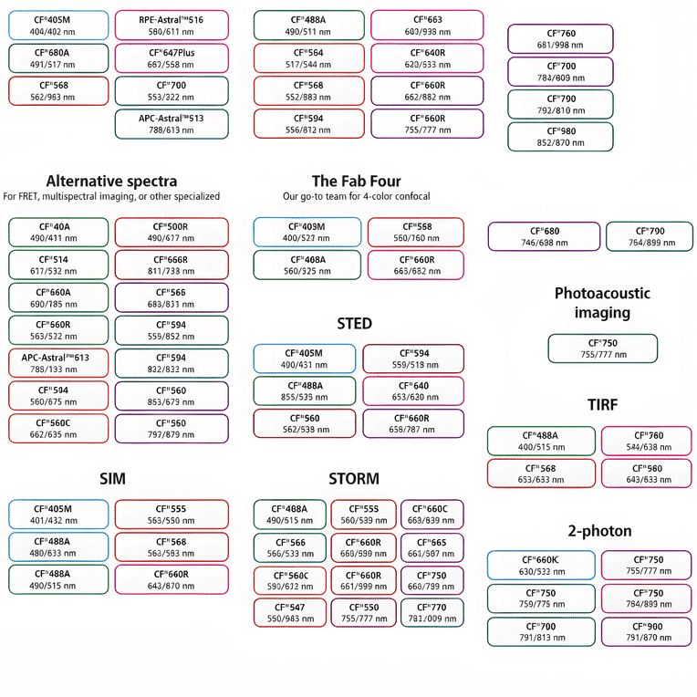
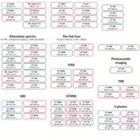
This chart summarizes commonly used CF® dyes grouped by their suitability for specific imaging modalities, including alternative spectra applications, four-color confocal imaging, near-infrared western blotting, photoacoustic imaging, STED, SIM, STORM, TIRF, and two-photon microscopy. Each dye is shown with its characteristic excitation and emission wavelengths (nm), providing a practical reference for selecting spectrally compatible dyes and optimizing multicolor experimental design across a range of fluorescence techniques.
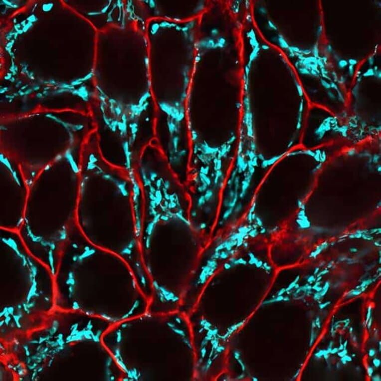
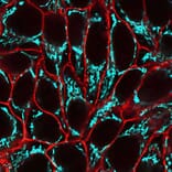
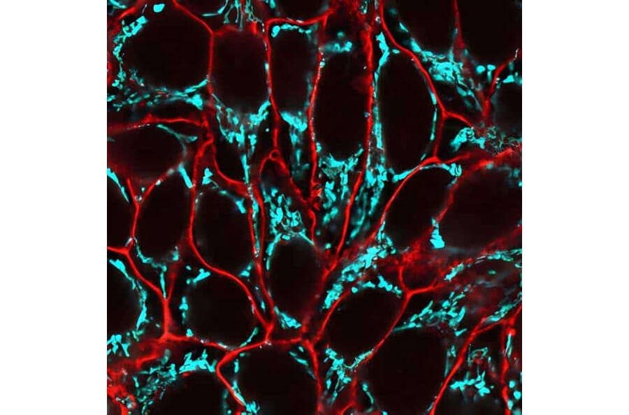
HeLa cells labeled with CellBrite® Fix 555 to stain the plasma membrane (red), fixed, and subsequently stained with CF®640R–conjugated anti-mitochondrial marker antibody (clone 113-1) to label mitochondria (cyan).
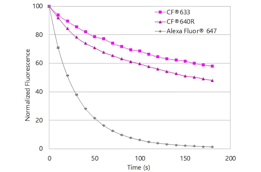
CF®640R exhibits substantially greater photostability than Alexa Fluor® 647. Jurkat cells were stained with mouse anti-CD3 primary antibody followed by goat anti-mouse secondary antibodies conjugated to the indicated dyes. Cells were continuously illuminated using a mercury arc lamp with a Cy®5 filter cube, with images acquired every 15 seconds for 5 minutes. Fluorescence intensity was normalized to the initial time point.
High-resolution STED imaging of cytoskeletal microtubules in U2OS cells. Tubulin filaments were immunolabeled with DM1A and visualized using a CF®640R-conjugated anti-mouse secondary antibody. Images were captured on a STELLARIS 8 STED FALCON system (Leica Microsystems GmbH, Germany).