Unconjugated
In vitro recapitulation of an organotypic stromal environment, enabling efficient angiogenesis, is crucial to investigate and possibly improve vascularization in regenerative medicine. Our study aims at engineering the complexity of a vascular milieu including multiple cell-types, a stromal extracellular matrix (ECM), and molecular signals. For this purpose, the human adipose stromal vascular fraction (SVF), composed of a heterogeneous mix of pericytes, endothelial/stromal progenitor cells, was cultured under direct perfusion flow on three-dimensional (3D) collagen scaffolds. Perfusion culture of SVF-cells reproducibly promoted in vitro the early formation of a capillary-like network, embedded within an ECM backbone, and the release of numerous pro-angiogenic factors. Compared to static cultures, perfusion-based engineered constructs were more rapidly vascularized and supported a superior survival of delivered cells upon in vivo ectopic implantation. This was likely mediated by pericytes, whose number was significantly higher (4.5-fold) under perfusion and whose targeted depletion resulted in lower efficiency of vascularization, with an increased host foreign body reaction. 3D-perfusion culture of SVF-cells leads to the engineering of a specialized milieu, here defined as an angiogenic niche. This system could serve as a model to investigate multi-cellular interactions in angiogenesis, and as a module supporting increased grafted cell survival in regenerative medicine.
To investigate the biocompatibility of the biomaterial, polylactic acid (PLA) with stem cells from human exfoliated deciduous teeth (SHED) and its induction of mineralization as a type of scaffold material. To determine the impacts of biomaterial PLA on proliferation and mineralization of SHED, the expression of surface molecules of SHED isolated and cultured in vitro was detected by flow cytometry. In addition, cell proliferation was measured using MTT and Edu assays, and the evaluation of mineralized differentiation was performed using Alizarin Red S staining. In addition, the expression levels of osteogenic marker genes were measured by reverse transcription-quantitative polymerase chain reaction (RT-qPCR) and western blot analysis. SHED were successfully isolated and identified. The MTT and Edu results indicated that the proliferation of SHED cultured in PLA and normal medium was not significantly different. The Alizarin Red S staining demonstrated that the mineralization capability was significantly higher in the SHED that were cultured in PLA medium. Furthermore, RT-qPCR and western blot analyses indicated that the expression levels of osteogenic marker genes were higher in the SHED cultured in PLA medium. These results suggested that PLA possesses good biocompatibility with SHED and may effectively induce the mineralization of SHED and serve as a scaffold material.


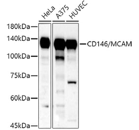
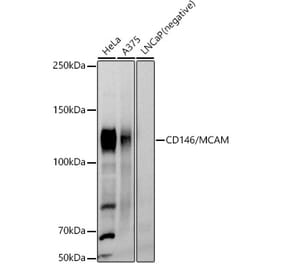
![Western Blot - Anti-CD146 Antibody [ARC1708] (A308820) - Antibodies.com](https://cdn.antibodies.com/image/catalog/308/A308820_1.jpg?profile=product_alternative)
![Immunohistochemistry - Anti-CD146 Antibody [MUC18/1130] (A249293) - Antibodies.com](https://cdn.antibodies.com/image/catalog/249/A249293_1.jpg?profile=product_alternative)
![Immunohistochemistry - Anti-CD146 Antibody [MUC18/1130] - BSA and Azide free (A252473) - Antibodies.com](https://cdn.antibodies.com/image/catalog/252/A252473_1.jpg?profile=product_alternative)
![Immunohistochemistry - Anti-CD146 Antibody [MCAM/1101] (A249290) - Antibodies.com](https://cdn.antibodies.com/image/catalog/249/A249290_1.jpg?profile=product_alternative)
![Immunohistochemistry - Anti-CD146 Antibody [MCAM/1101] - BSA and Azide free (A252470) - Antibodies.com](https://cdn.antibodies.com/image/catalog/252/A252470_1.jpg?profile=product_alternative)

![Immunohistochemistry - Anti-CD146 Antibody [C146/634] - BSA and Azide free (A252471) - Antibodies.com](https://cdn.antibodies.com/image/catalog/252/A252472_1.jpg?profile=product_alternative)
![Immunohistochemistry - Anti-CD146 Antibody [MCAM/3046] (A249295) - Antibodies.com](https://cdn.antibodies.com/image/catalog/249/A249295_1.jpg?profile=product_alternative)
![Immunohistochemistry - Anti-CD146 Antibody [MCAM/3046] - BSA and Azide free (A252475) - Antibodies.com](https://cdn.antibodies.com/image/catalog/252/A252475_1.jpg?profile=product_alternative)