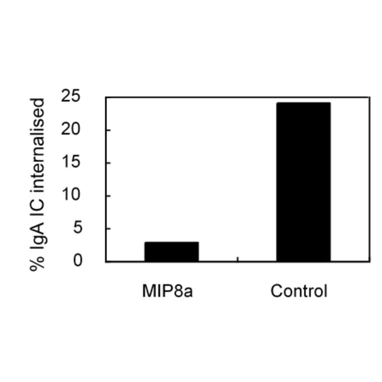
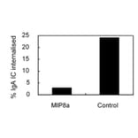
Unconjugated
FcalphaR, the Fc receptor for IgA, is essential for IgA-mediated immune responses. Previous studies have shown that IgA and IgA immune complexes can be rapidly endocytosed by FcalphaR. However, the underlying mechanism remains unclear. Here, we investigated the endocytic pathway of FcalphaR in monocytic cell line, U937, that naturally express FcalphaR and in transfected Chinese hamster ovary (CHO), COS-7 and Hela cells. By using selective chemical inhibitors of different endocytic pathways, overexpression of dominant-negative mutants of Eps15 and knockdown of clathrin heavy chain (CHC) via RNA interference, we demonstrated that endocytosis of FcalphaR was through a clathrin-mediated pathway. The endocytosed FcalphaR went into Rab5- and Rab11-positive endosomes. However, endocytosis of FcalphaR could not be blocked by a dominant-negative mutant of Rab5. We also demonstrated that endocytosis of FcalphaR was dynamin-dependent by overexpressing a dominant-negative mutant of dynamin. The potential endocytic motif for FcalphaR was also examined. Unexpectedly, we found that the entire cytoplasmic domain of FcalphaR was not required for the endocytic process of FcalphaR. We conclude that endocytosis of FcalphaR is clathrin- and dynamin-dependent, but is not regulated by Rab5, and the endocytic motif is not located in the cytoplasmic domain of FcalphaR.
The human IgA Fc receptor (FcalphaR, CD89) plays an important role in host defence against invading pathogens. To study the properties of the receptor, 12 MoAbs, namely, MIP7c, MIP8a, MIP9a, MIP10c, MIP11c, MIP14b, MIP15b, MIP38c, MIP59c, MIP65c, MIP68b and MIP71a, were generated. The inhibitory effects of the antibodies on FcalphaR functions were tested. Three of the antibodies, MIP7c, MIP8a and MIP59c, were able to block up to 90% of soluble FcalphaR binding to IgA-coated beads and 70-80% of neutrophil phagocytosis of IgA immune complexes (IC). MIP8a could also inhibit IgA IC-induced neutrophil lactoferrin release, while cross-linking of FcalphaR with MIP8a and anti-mouse IgG could elicit neutrophil lactoferrin release. However, IgA IC-induced lactoferrin release required both extracellular calcium and magnesium, whereas MIP8a-induced release did not require extracellular magnesium and only partially required extracellular calcium. In addition, the time course of IgA IC-induced lactoferrin release was slow. Lactoferrin was not detectable if the incubation time was less than 0.5 h. In contrast, MIP8a-induced lactoferrin release was fast. Lactoferrin could be detected within 5 min of incubation. Therefore, neutrophil lactoferrin release induced by IgA IC differed from that induced by cross-linking of FcalphaR with MIP8a.