A synthetic peptide corresponding to residues in Human CD9.
Host
Rabbit
Clonality
Polyclonal
Conjugate
Unconjugated
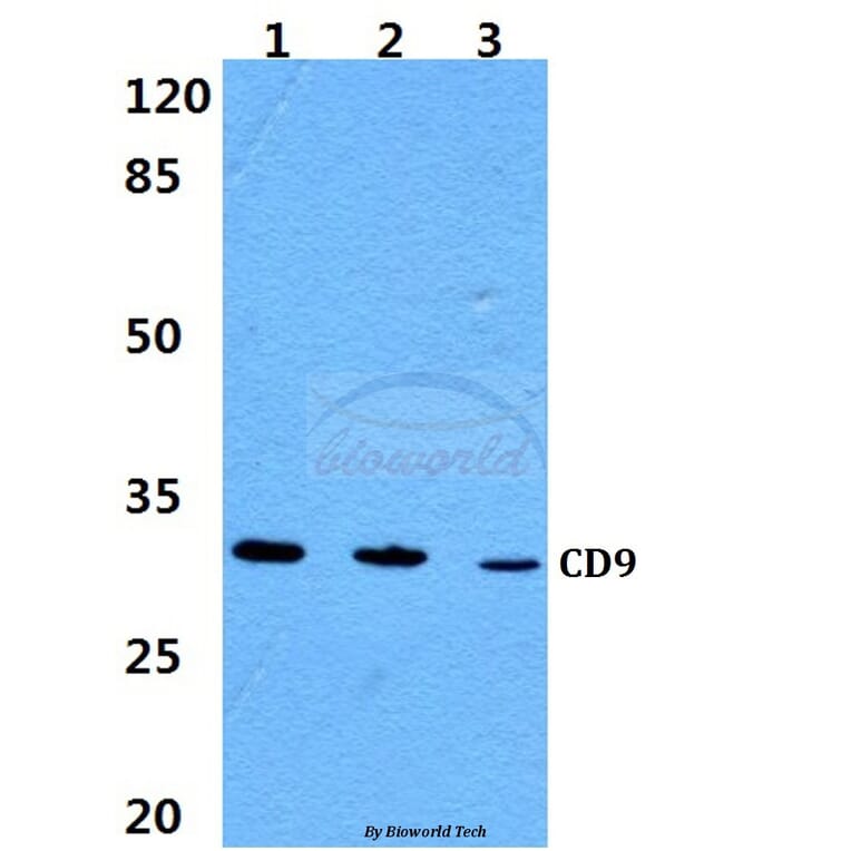
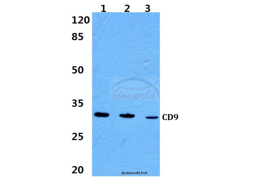
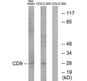
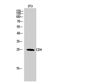
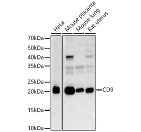
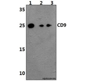
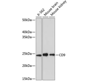
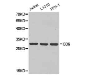
Molecular Weight
~ 30 kDa
Purity
The antibody was affinity-purified from rabbit antiserum by affinity-chromatography using epitope-specific immunogen and the purity is > 95% (by SDS-PAGE).
Product Form
1 mg/ml in Phosphate buffered saline (PBS) with 0.05% sodium azide, approx. pH 7.2.


![Flow Cytometry - Anti-CD9 Antibody [MEM-61] (A86089)](https://cdn.antibodies.com/image/catalog/86/A86089_1.jpg?profile=product_alternative)

![Flow Cytometry - Anti-CD9 Antibody [IVA50] (A86286)](https://cdn.antibodies.com/image/catalog/86/A86286_1.jpg?profile=product_alternative)
![Flow Cytometry - Anti-CD9 Antibody [MEM-61] - BSA and Azide free (A86091)](https://cdn.antibodies.com/image/catalog/86/A86091_1.jpg?profile=product_alternative)
![Flow Cytometry - Anti-CD9 Antibody [EM-04] (A86315)](https://cdn.antibodies.com/image/catalog/86/A86315_1.jpg?profile=product_alternative)
![SDS-PAGE - Anti-CD9 Antibody [Research Grade Biosimilar] - Low endotoxin, Azide free (A323956) - Antibodies.com](https://cdn.antibodies.com/image/catalog/323/A323956_1.jpg?profile=product_alternative)