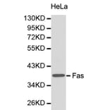
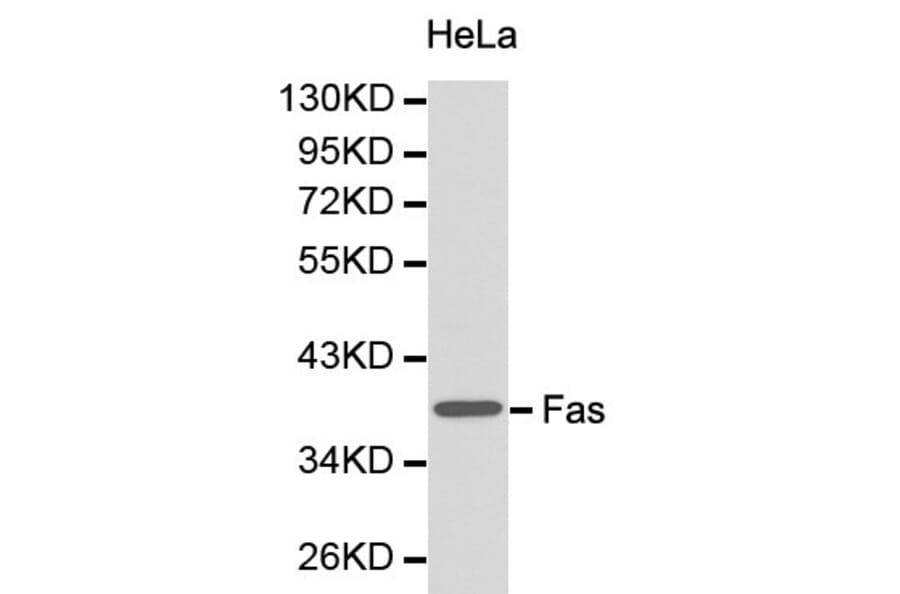
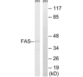
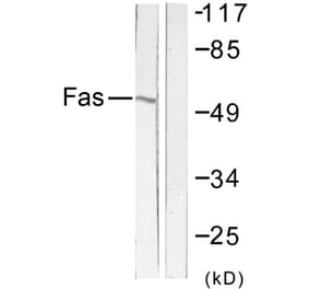
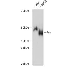
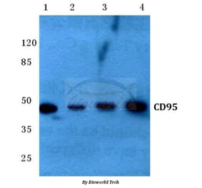
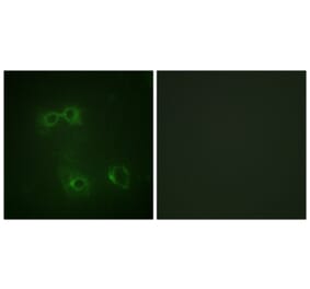
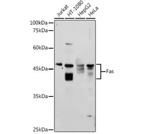
Unconjugated
Endothelial injury has been implicated in the pathogenesis of many cardiovascular diseases, including thrombotic disorders. Hyperin (quercetin-3-O-galactoside), a flavonoid compound and major bioactive component of the medicinal herb Apocynum venetum L., is commonly used to prevent endothelium dysfunction. However, its mode of action remains unclear. To the best of our knowledge, we have for the first time investigated the protective effect hyperin exerts against H2O2-induced injury in human endothelium-derived EA.hy926 cells using isobaric tags for relative and absolute quantitation (iTRAQ)‑based quantitative proteomic analysis. The results showed that H2O2 exposure induced alterations in the expression of 250 proteins in the cells. We noted that the expression of 52 proteins associated with processes such as cell apoptosis, cell cycle and cytoskeleton organization, was restored by hyperin treatment. Of the proteins differentially regulated following H2O2 stress, the anti-apoptotic protein, myeloid cell leukemia-1 (Mcl-1), and the pro-apoptotic protein, BH3-interacting domain death agonist (Bid), exhibited marked changes in expression. Hyperin increased Mcl-1 expression and decreased that of Bid in a dose-dependent manner. In addition, flow cytometric analysis and western blot analysis of the apoptosis-related proteins, truncated BID (tBid), cleaved caspase-3, cleaved caspase-9, Fas, FasL and caspase-8, demonstrated that the rate of apoptosis and the pro-apoptotic protein levels were decreased by hyperin pre‑treatment. In the present study we demonstrate that hyperin effectively prevents H2O2‑induced cell injury by regulating the Mcl‑1‑ and Bid-mediated anti‑apoptotic mechanism, suggesting that hyperin is a potential candidate for use in the treatment of thrombotic diseases.
Actinomycin D (Act D), a well known of clinical antitumor drug, has been used for the treatment of some highly malignant tumors, however, the clinical application was limited by its extreme cytotoxicity. In the present study, we reported that methylated actinomycin D (mAct D), a novel actinomycin D analog isolated from Streptomyces sp. KLBMP 2541 in our previous study, could not only exert stronger inhibitory effects on several human cancer cells than Act D in dose- and time-dependent manner at ng concentrations, especially on HepG2 cells, but also lower cytotoxicity in normal cells (HL-7702). Base on these results, HepG2 cells were treated for further study to illustrate the potential mechanism of mAct D. The results of nuclei morphology examination, DNA fragmentation detection, sub-G1 analysis, annexin V-FITC/PI staining and activation of caspase-3 indicated mAct D significantly induced HepG2 cells apoptosis. Semiquantitative RT-PCR and Western blot analysis revealed that mAct D induced apoptosis in HepG2 cells through mitochondria-dependent pathway by increasing levels of caspase-9, Bax, Bak while decreasing levels of Bcl-2, Bid, and Fas-dependent pathway by increasing levels of Fas, FasL, FADD, and caspase-8. Subsequently, pretreatment with specific inhibitor of caspase-8 Z-LEHD-FMK and caspase-9 Z-LEHD-FMK significantly attenuated caspase-3 activity, the cleavage of caspase-3 and PARP, meanwhile increased the cell viability. In addition, p53 and mitochondrial transcription factor A (mtTFA) were also upregulated. Taken together, ng concentrations mAct D induces the apoptosis of HepG2 through Fas- and mitochondria-mediated pathway and presents a potential novel alternative agent for the treatment of human hepatic carcinoma.