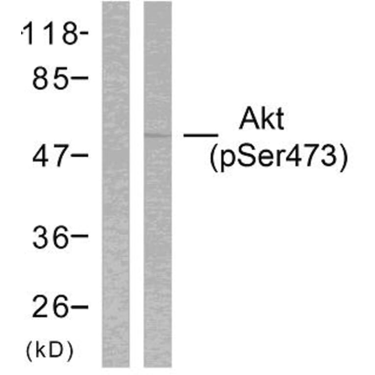
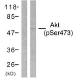
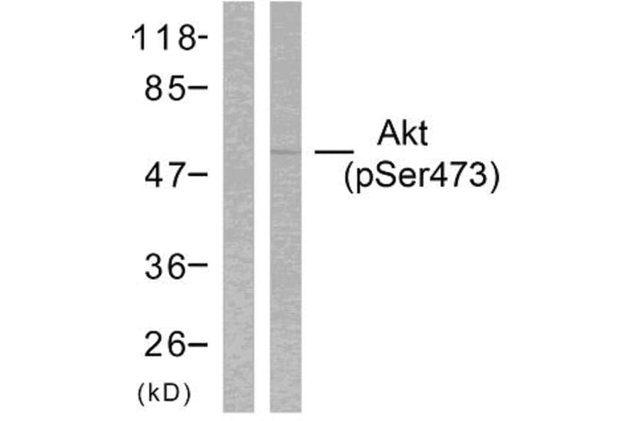
Western Blot - Anti-Akt (phospho Ser473) Antibody (A94682)
Western blot analysis of lysates from HeLa cells treated with heat shock using Anti-Akt (phospho Ser473) Antibody. The left hand lane represents a negative control, where the antibody is blocked by the immunising peptide.
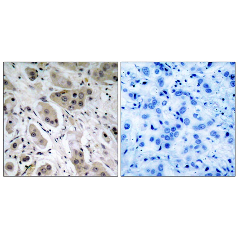
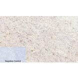
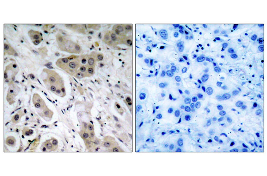
Immunohistochemical analysis of paraffin-embedded human breast carcinoma using Anti-Akt (phospho Ser473) Antibody. The right hand panel represents a negative control, where the antibody was pre-incubated with the immunising peptide.
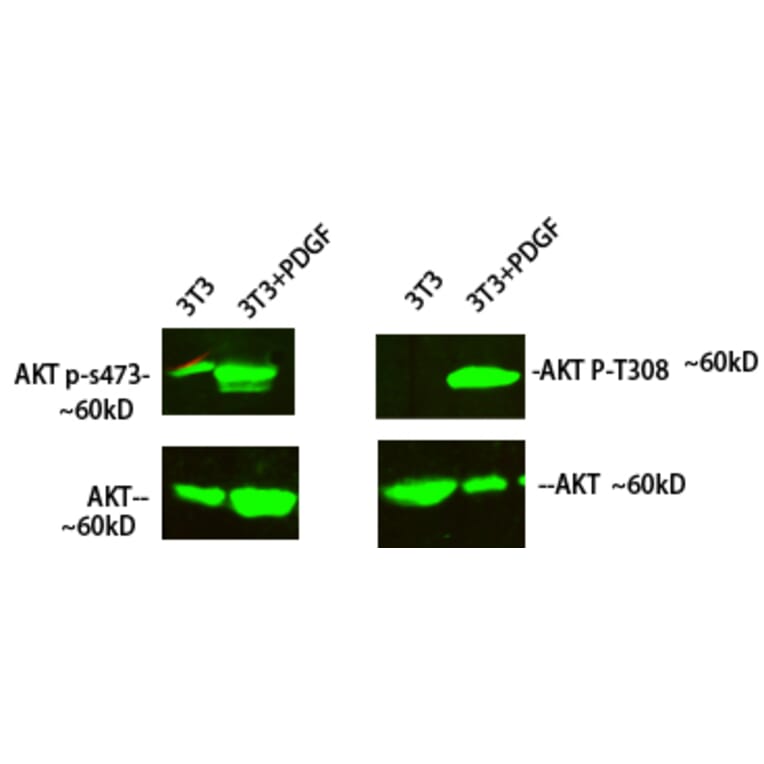
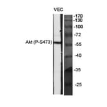
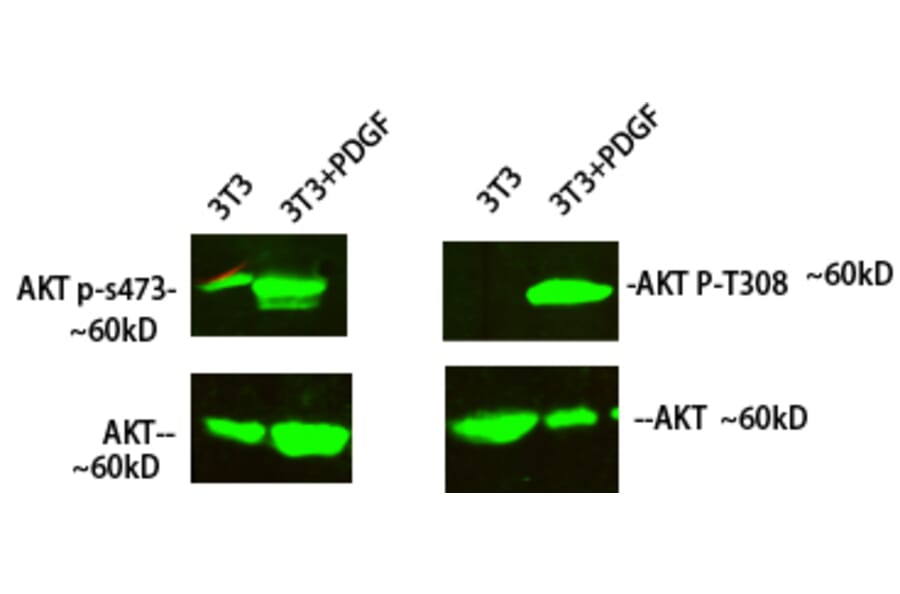
Western Blot - Anti-Akt (phospho Ser473) Antibody (A94682)
Western blot analysis of 3T3 cells treated with PDGF using Anti-Akt (phospho Ser473) Antibody at 1:1,000 (4°C overnight). Goat Anti-Rabbit IgG (IRDye 800) was used as a secondary at 1:5,000 (25°C, 1 hour).
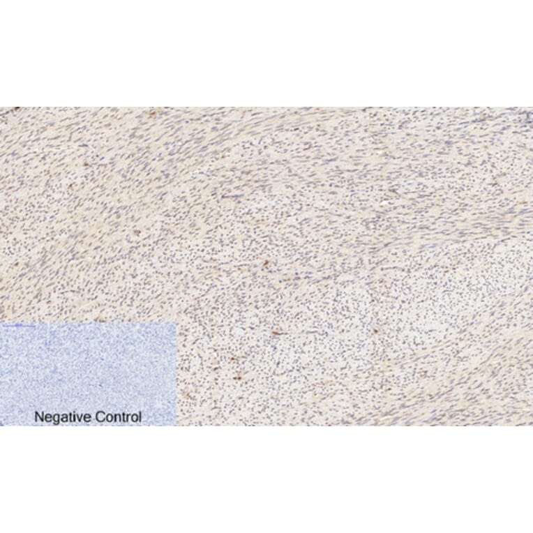
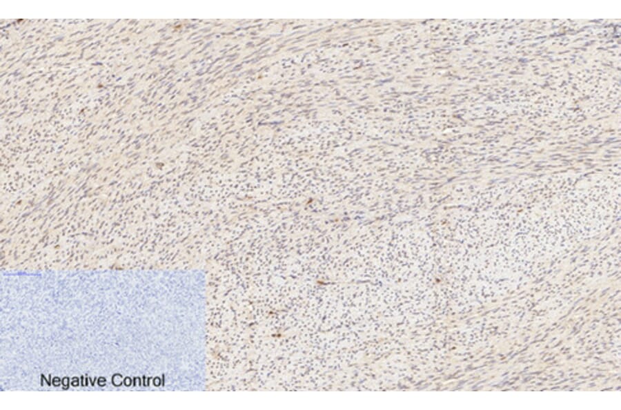
Immunohistochemical analysis of paraffin-embedded human uterus tissue using Anti-Akt (phospho Ser473) Antibody at 1:200 (4°C overnight). Negative control was secondary antibody only.
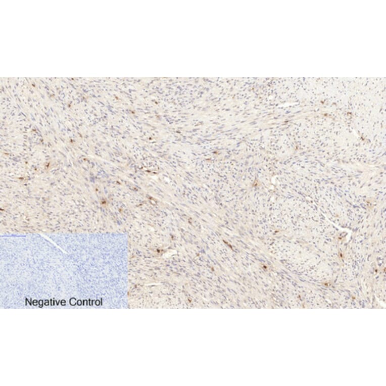
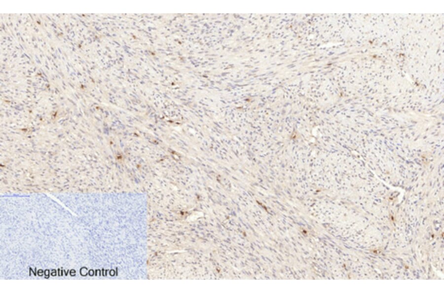
Immunohistochemical analysis of paraffin-embedded human uterus cancer tissue using Anti-Akt (phospho Ser473) Antibody at 1:200 (4°C overnight). Negative control was secondary antibody only.
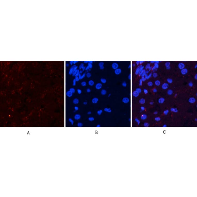
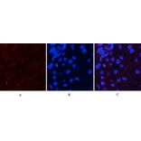
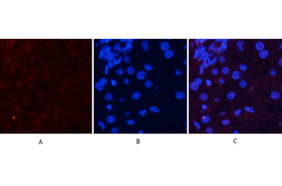
Immunofluorescence analysis of rat liver tissue using Anti-Akt (phospho Ser473) Antibody (red) at 1:200 (4°C overnight). Cy3 labelled secondary antibody was used at 1:300 (RT 50min). Panel A: Target. Panel B: DAPI. Panel C: Merge.
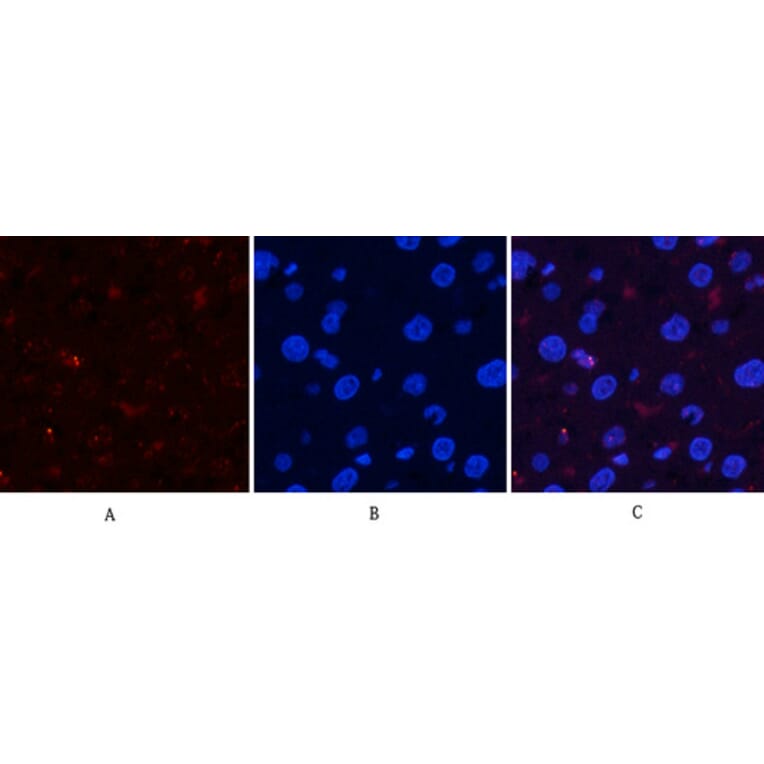
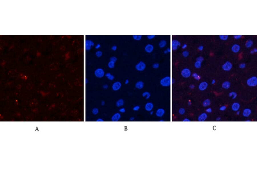
Immunofluorescence analysis of rat liver tissue using Anti-Akt (phospho Ser473) Antibody (red) at 1:200 (4°C overnight). Cy3 labelled secondary antibody was used at 1:300 (RT 50min). Panel A: Target. Panel B: DAPI. Panel C: Merge.