Mouse monoclonal [TU-06] antibody to beta Tubulin.
Specificity
The antibody recognizes an epitope (aa 81-95) on phylogenetically conserved N-terminal structural domain of beta-tubulin (recognizes all beta-tubulin isoforms) in various species.
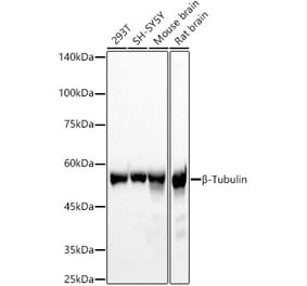
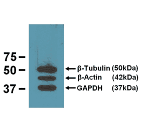
Anti-beta Tubulin Antibody [TU-06] (A86335) works in WB application under reducing conditions on RIPA cell extracts. Western blotting analysis was performed on whole cell extracts (RIPA lysis buffer) of HeLa, HEK 293, ESS-1 and Jurkat cell lines mixed and heated (100°C, 5 min) with reducing (2-mercaptoethanol) or non-reducing SDS-loading buffer. Samples were resolved using 12% Tris-glycine SDS gel electrophoresis. Nitrocellulose membrane blot was probed simultaneously with mouse IgM monoclonal antibody TU-06 (1 µg/ml) and mouse IgG1 anti-GAPDH monoclonal antibody FF26A (1 µg/ml) used as the loading control. Subclass-specific secondary antibodies IRDye 680RD Goat-anti-Mouse IgM (red) and IRDye 800CW Goat-anti-Mouse IgG (green) were used for multiplex fluorescent Western blot detection. Alpha-tubulin was detected at ~50 kDa in all tested cell lines under reducing, but not under non- reducing conditions. Using RIPA lysis buffer in combination with non-reducing conditions is not suitable for Anti-beta Tubulin Antibody [TU-06] (A86335).
Immunocytochemistry staining of 3T3 mouse embryonal fibroblast cell line using Anti-beta Tubulin Antibody [TU-06] (A86335), (detection by Goat anti-mouse IgM Cy®5). Nucleus is stained with DAPI (blue).
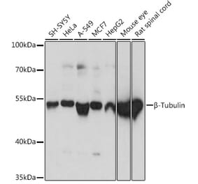
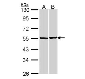
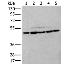
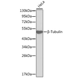
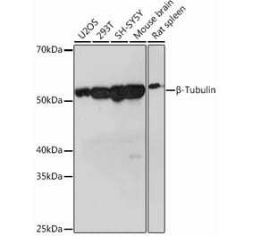
Western blotting analysis of human beta-tubulin using Anti-beta Tubulin Antibody [TU-06] (A86335) on lysates (50 mM TRIS-Cl pH 6.8, 4M UREA, 4% SDS) of various cell lines under non-reducing and reducing conditions. Nitrocellulose membrane was probed with 2 µg/ml of mouse anti-beta-tubulin monoclonal antibody followed by IRDye800-conjugated anti-mouse secondary antibody. A specific band was detected for beta-tubulin at approximately 54 kDa.
Publishing research using Anti-beta Tubulin Antibody [TU-06] (A86335)? Please let us know so that we can list the citation on this page.


![WB - Anti-beta Tubulin Antibody [TU-06] (A86335)](https://cdn.antibodies.com/image/catalog/86/A86335_1.jpg?profile=product_top)
![ICC - Anti-beta Tubulin Antibody [TU-06] (A86335)](https://cdn.antibodies.com/image/catalog/86/A86335_2.jpg?profile=product_top)
![IHC - Anti-beta Tubulin Antibody [TU-06] (A86335)](https://cdn.antibodies.com/image/catalog/86/A86335_3.jpg?profile=product_top)
![WB - Anti-beta Tubulin Antibody [TU-06] (A86335)](https://cdn.antibodies.com/image/catalog/86/A86335_4.jpg?profile=product_top)
![WB - Anti-beta Tubulin Antibody [TU-06] (A86335)](https://cdn.antibodies.com/image/catalog/86/A86335_1.jpg?profile=product_top_thumb)
![ICC - Anti-beta Tubulin Antibody [TU-06] (A86335)](https://cdn.antibodies.com/image/catalog/86/A86335_2.jpg?profile=product_top_thumb)
![IHC - Anti-beta Tubulin Antibody [TU-06] (A86335)](https://cdn.antibodies.com/image/catalog/86/A86335_3.jpg?profile=product_top_thumb)
![WB - Anti-beta Tubulin Antibody [TU-06] (A86335)](https://cdn.antibodies.com/image/catalog/86/A86335_4.jpg?profile=product_top_thumb)
![WB - Anti-beta Tubulin Antibody [TU-06] (A86335)](https://cdn.antibodies.com/image/catalog/86/A86335_1.jpg?profile=product_image)
![ICC - Anti-beta Tubulin Antibody [TU-06] (A86335)](https://cdn.antibodies.com/image/catalog/86/A86335_2.jpg?profile=product_image)
![IHC - Anti-beta Tubulin Antibody [TU-06] (A86335)](https://cdn.antibodies.com/image/catalog/86/A86335_3.jpg?profile=product_image)
![WB - Anti-beta Tubulin Antibody [TU-06] (A86335)](https://cdn.antibodies.com/image/catalog/86/A86335_4.jpg?profile=product_image)
![Immunofluorescence - Anti-beta Tubulin Antibody [1B12] (A85428) - Antibodies.com](https://cdn.antibodies.com/image/catalog/85/A85428_1.jpg?profile=product_alternative)
![WB - Anti-beta Tubulin Antibody [TU-13] (A86730)](https://cdn.antibodies.com/image/catalog/86/A86730_1.jpg?profile=product_alternative)

![Immunohistochemistry - Anti-beta Tubulin Antibody [4E4] (A85429) - Antibodies.com](https://cdn.antibodies.com/image/catalog/85/A85429_1.jpg?profile=product_alternative)