

Unconjugated
Expression of the avß6 integrin is upregulated on multiple solid epithelial tumors while this integrin is largely absent on the healthy human epithelium. In the present study, we sought to compare the in vivo biodistributions of three avß6-selective agents that could be used for the positron emission tomography (PET)-based detection of malignancies expressing this integrin. A streptavidin-coupled avß6-binding peptide (streptavidin-A20), an anti-avß6 monoclonal antibody (620W.7) and an avß6-targeted protein, derived from a modified adenovirus 5 receptor-binding domain (Ad5.KO1.A20), were radiolabeled with [89Zr] and intravenously injected into mice bearing bilateral avß6-positive and -negative melanoma xenografts. Micro-PET imaging was performed at 0.33, 24, 48, 72, and (620W.7 group only) 144 h post-injection. Streptavidin-A20-based radiotracers preferentially accumulated in avß6-positive tumors, achieving >3.4:1 A375-ß6:A375 activity ratios between 24 and 72 h post-injection, with off-target uptake restricted to the kidneys. 620W.7 exhibited roughly 2-fold greater accumulation in A375-ß6 than A375 tumors, with lower levels of renal retention and greater activity detected in the liver and spleen. Despite having demonstrated selective binding to avß6-expressing cells in vitro, the Ad5.KO1.A20 protein failed to selectively traffic to tumors expressing this integrin in vivo. Nevertheless, streptavidin-A20 and 620W.7 demonstrated promise as avß6-selective agents for in vivo applications.
Mycobacteria, including the tuberculosis pathogen Mycobacterium tuberculosis, are enclosed by a highly complex cell envelope with an outer membrane, or mycomembrane, which provides extraordinary protection from antibiotics and other stresses. The inner leaflet of the mycomembrane consists of arabinogalactan-linked mycolate (AGM), which is an enormous glycoconjugate comprising mycolic acids esterified to terminal D-arabinofuranosyl residues of an underlying arabinogalactan-peptidoglycan complex, also referred to as the mycoloyl-arabinogalactan-peptidoglycan (mAGP) complex. Whereas AGM biosynthesis is comparatively well characterized, less is known about AGM degradation by endogenous or exogenous factors. To facilitate studies on AGM breakdown by hydrolytic enzymes, here we synthesized fluorescence resonance energy transfer (FRET)-based mono- and disaccharide probes that mimic fragments of AGM and are designed to fluoresce upon cleavage. We devised a synthetic route that established the glycolipid core with the desired regio- and stereochemistry and allowed late-stage selective functionalization of the core with a FRET pair. Our data show that the intact FRET-AGM probes exist in a fluorescence-quenched state, but when exposed to lysin B (LysB), an AGM-degrading mycobacteriophage hydrolase with therapeutic relevance, the probes were activated through lipid ester hydrolysis, thereby generating fluorescence signal. FRET-AGM probes were activated by known mycomembrane glycolipid hydrolases, but not by several other types of hydrolases, demonstrating specificity. FRET-AGM probes may be useful in the future for identifying novel AGM hydrolases and quantitatively monitoring the activity of AGM hydrolases, which could provide insights into mycomembrane degradative processes and aid in tuberculosis therapeutic development.

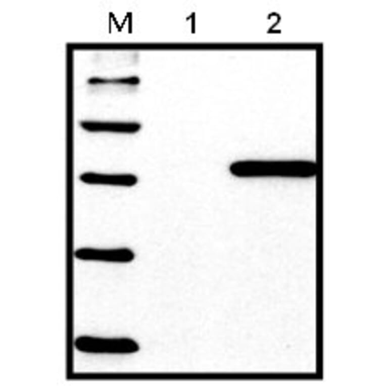
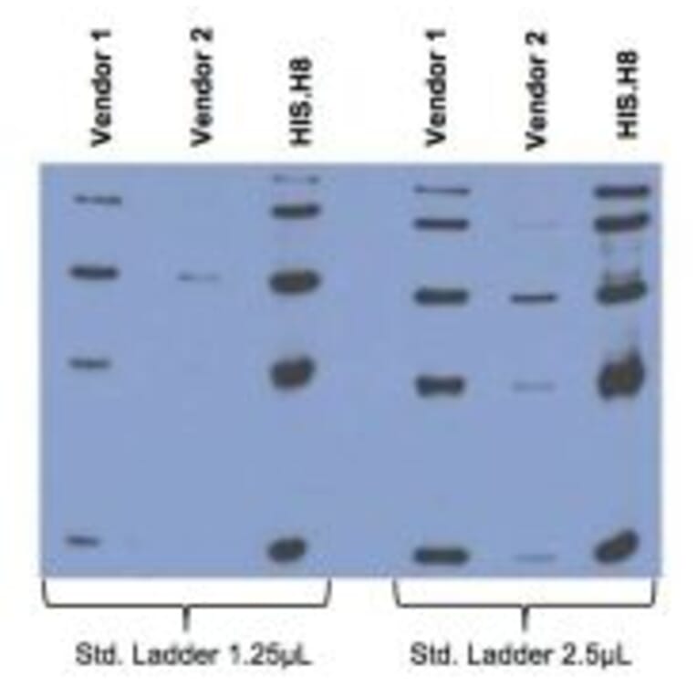
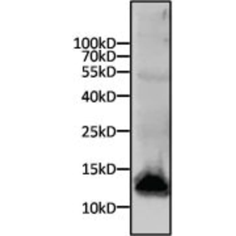
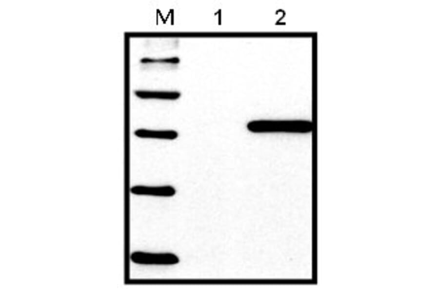
![TEM - Anti-His Tag Antibody [HIS.H8] - Antibodies.com](https://cdn.antibodies.com/image/catalog/85/A85277_140498838.jpg?profile=product_top)
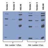
![TEM - Anti-His Tag Antibody [HIS.H8] - Antibodies.com](https://cdn.antibodies.com/image/catalog/85/A85277_240498838.jpg?profile=product_top)
![TEM - Anti-His Tag Antibody [HIS.H8] - Antibodies.com](https://cdn.antibodies.com/image/catalog/85/A85277_340498838.jpg?profile=product_top)
![TEM - Anti-His Tag Antibody [HIS.H8] - Antibodies.com](https://cdn.antibodies.com/image/catalog/85/A85277_140498838.jpg?profile=product_top_thumb)
![TEM - Anti-His Tag Antibody [HIS.H8] - Antibodies.com](https://cdn.antibodies.com/image/catalog/85/A85277_240498838.jpg?profile=product_top_thumb)
![TEM - Anti-His Tag Antibody [HIS.H8] - Antibodies.com](https://cdn.antibodies.com/image/catalog/85/A85277_340498838.jpg?profile=product_top_thumb)

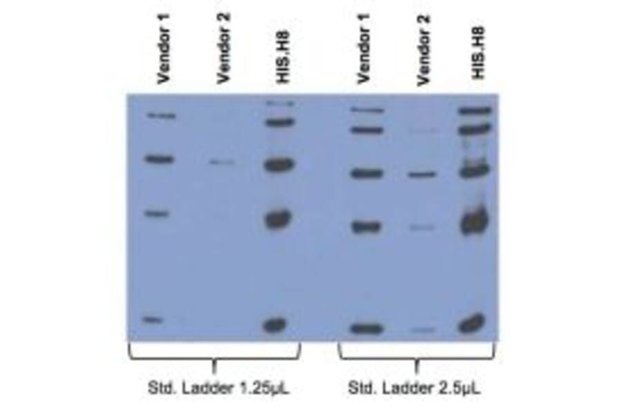
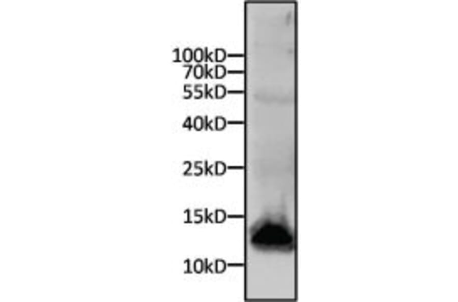
![TEM - Anti-His Tag Antibody [HIS.H8] - Antibodies.com](https://cdn.antibodies.com/image/catalog/85/A85277_140498838.jpg?profile=product_image)
![TEM - Anti-His Tag Antibody [HIS.H8] - Antibodies.com](https://cdn.antibodies.com/image/catalog/85/A85277_240498838.jpg?profile=product_image)
![TEM - Anti-His Tag Antibody [HIS.H8] - Antibodies.com](https://cdn.antibodies.com/image/catalog/85/A85277_340498838.jpg?profile=product_image)