Luca Murru, Italian National Research Council
Verified Customer
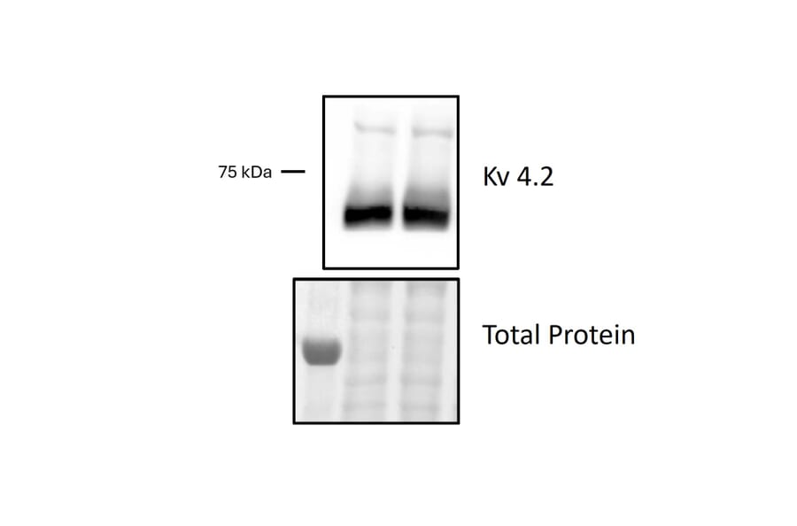
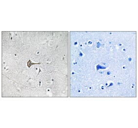
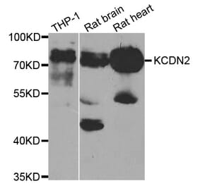
Applications: Western Blot
Species: Mouse
Sample: Mouse brain tissue (hippocampus)
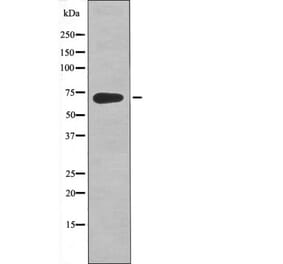
Experimental Conditions: WB: Mouse brain tissue lysate prepared in 1% SDS, 10 mM EDTA pH 8.0, 50 mM Tris pH 8.0 with protease and phosphatase inhibitors; 25 µg total protein loaded per lane; samples supplemented with Laemmli buffer and heated at 95°C for 5 min; 7% SDS-PAGE; nitrocellulose membrane; wet transfer in 25 mM Tris, 192 mM glycine, 20% methanol for 90 min at 80 V, 4°C; transfer verified by Ponceau S staining; blocking in 5% BSA/TBST for 1 h RT; primary antibody 1:1000 overnight at 4°C; washes 3×10 min in TBST; secondary antibody Jackson ImmunoResearch 111-035-144, 1:10000 in 5% milk/TBST for 1 h RT; detection by ECL.
Dilution: Primary: 1:1000; Secondary: 1:10000
Results Summary: A prominent band was detected at approximately 71 kDa in mouse hippocampal lysate, consistent with the expected molecular weight of KV4.2. The signal was reproducible between lanes, although moderate background staining and some non-specific bands were observed.
Comments: The antibody detected KV4.2 under the tested conditions; further optimisation of blocking conditions or antibody dilution may reduce background and improve specificity.