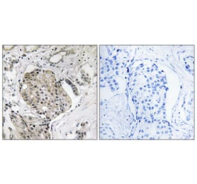
ILKNUR SUR ERDEM, Centre for Medicines Discovery (CMD), University of Oxford
Verified Customer
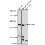
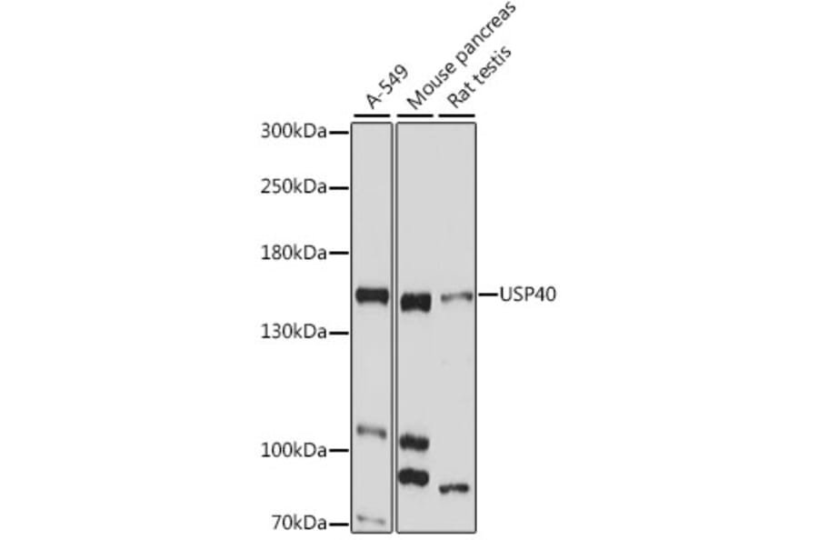
Applications: Western Blot
Species: Human
Sample: Human cell line
Experimental Conditions: Western blot analysis was performed using human cell line lysates prepared in Co-IP lysis buffer (20 mM HEPES pH 8.0, 150 mM NaCl, 0.2% NP-40, 10% glycerol) supplemented with fresh protease and phosphatase inhibitors. Cells were treated with DMSO, bafilomycin A1 (100 nM), and MG132 (100 µM) for 5 h prior to lysis. Lysates were clarified at 14,000×g for 10 min at 4 °C, quantified by BCA assay, and 60 µg total protein per lane was resolved by SDS-PAGE. Proteins were transferred to a PVDF membrane by wet blotting at constant 400 mA for 60 min. Membranes were blocked in 4% BSA in PBS-T for 1 h at room temperature and incubated with primary antibody overnight at 4 °C. Detection was performed using LI-COR near-infrared fluorescence imaging.
Dilution: Primary: 1:1000 (PBS-T + 1% BSA); Secondary: LI-COR IRDye 680RD Goat anti-Rabbit IgG (H+L), 926-68071, 1:10000 for 1 h.
Results Summary: Successful detection of USP40 in immunoprecipitation input extracts from wild-type and protein X knockout cell lines using the Anti-USP40 Antibody (A88240). A distinct band is observed at the expected molecular weight with minimal background signal, indicating specific and reliable target recognition under the described conditions.
Comments: A detailed protocol was also provided by the customer.