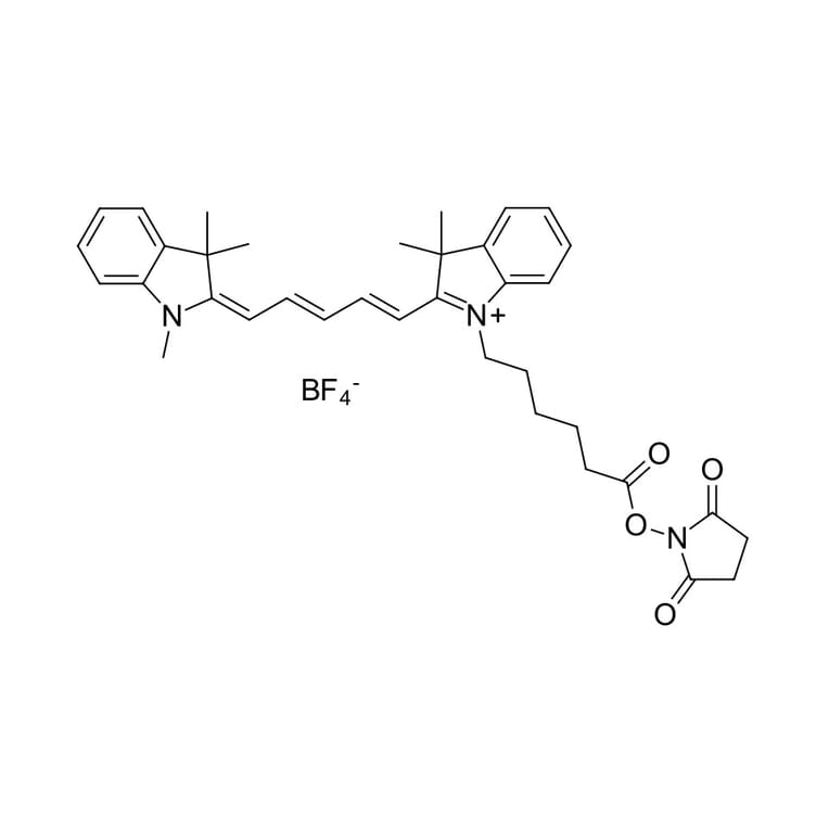
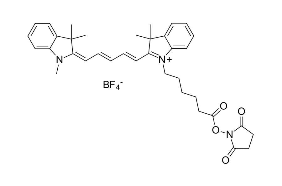
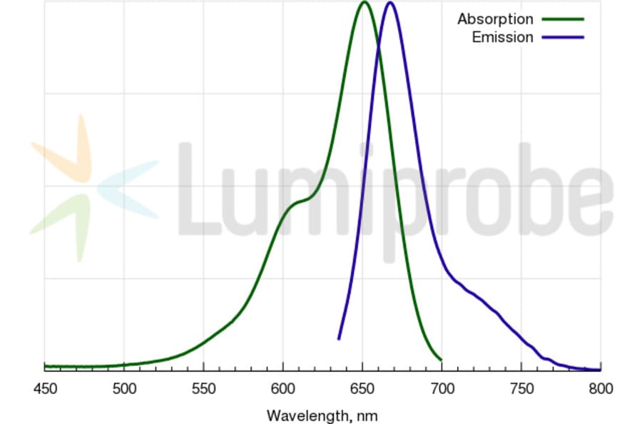
Natural killer (NK) cells are key innate immunity effectors that combat viral infections and control several cancer types. For their immune function, human NK cells rely largely on five different cytotoxic proteases, called granzymes (A/B/H/K/M). Granzyme B (GrB) initiates at least three distinct cell death pathways, but key aspects of its function remain unexplored because selective probes that detect its activity are currently lacking. In this study, we used a set of unnatural amino acids to fully map the substrate preferences of GrB, demonstrating previously unknown GrB substrate preferences. We then used these preferences to design substrate-based inhibitors and a GrB-activatable activity-based fluorogenic probe. We show that our GrB probes do not significantly react with caspases, making them ideal for in-depth analyses of GrB localization and function in cells. Using our quenched fluorescence substrate, we observed GrB within the cytotoxic granules of human YT cells. When used as cytotoxic effectors, YT cells loaded with GrB attacked MDA-MB-231 target cells, and active GrB influenced its target cell-killing efficiency. In summary, we have developed a set of molecular tools for investigating GrB function in NK cells and demonstrate noninvasive visual detection of GrB with an enzyme-activated fluorescent substrate.
Chitosan-based nanosystems have been described as interesting tools for antigen delivery and for enhancing the immunogenicity of nasally administered vaccines. As a possible vaccine delivery method, the chemical conjugation of chitosan nanocapsules with the Streptococcus pneumoniae cell membrane protein PsaA (pneumococcal surface adhesin A) is suggested here. The antigen PsaA, common to all pneumococcus serotypes, is expected to improve its uptake by immune cells and to activate specific T cells, generating an adaptive immune response against pneumococcus. With this aim, chitosan nanocapsules with thiol-maleimide conjugation between the polymer (chitosan) and the antigen (PsaA) were designed to enable the surface presentation of PsaA for immune cell recognition. Spherical-shaped particles, with a size of 266 ± 32 nm, positive charge of +30 ± 1 mV, and good stability profiles in simulated nasal fluids (up to 24 h) were achieved. PsaA association rates were three times higher compared with nanocapsules without covalent polymer-protein conjugation. Cytotoxicity studies in cell culture media showed non-toxic effect under 150 µg/mL concentration of nanocapsules, and subsequent studies on the maturation of immature dendritic cells in the presence of antigen-conjugated nanocapsules displayed peripheral blood mononuclear cell activation and lymphocyte differentiation after their presentation by dendritic cells. Secretion of TNFa following exposure to nanocapsules and the ability of nanocapsules to activate CD4 and CD8 T lymphocytes had also been studied. Antigen loaded nanocarrier uptake and presentation by professional presenting cells.